
Superior Frontal Sulcus
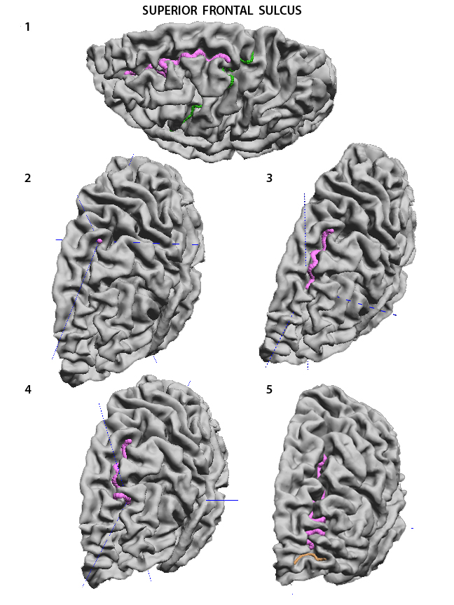
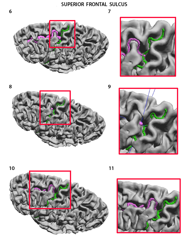
The Superior frontal Sulcus (SFS) separates the superior frontal from the middle frontal gyrus. It is seen best when looking at the dorsolateral surface of the hemisphere from above. It has a postero-anterior course parallel to the interhemispheric fissure (1). It starts at the preCS (1), with which it can merge or even cross, and it ends at the Fronto-Marginal Sulcus (see below) close to the frontal pole (5). It is very often interrupted and has very deep and sometimes long side branches. Drop the first point of the curve at the posterior end, immediately in front of the preCS (2), even if the SFS reaches the preCS or crosses it (see 6-11). Then proceed to the end of that segment (3). To jump over the gyrus interrupting the sulcus select no stickiness which helps direct the trace exactly where it should go (4), and go to the anterior end of the curve (5). When the SFS merges with the preCS as shown in 6, the curve cannot start at the exact point where the two sulci join. The curve on 6 (detail in 7) is wrong. Start the curve immediately above the preCS (8, detail in 9). Then proceed in the usual way. The final correct curve will be as in 10 (detail in 11). |
|
USC Biomedical Imaging Research Lab © 2009, designed by Dimitrios Pantazis |