
Circular Sulcus
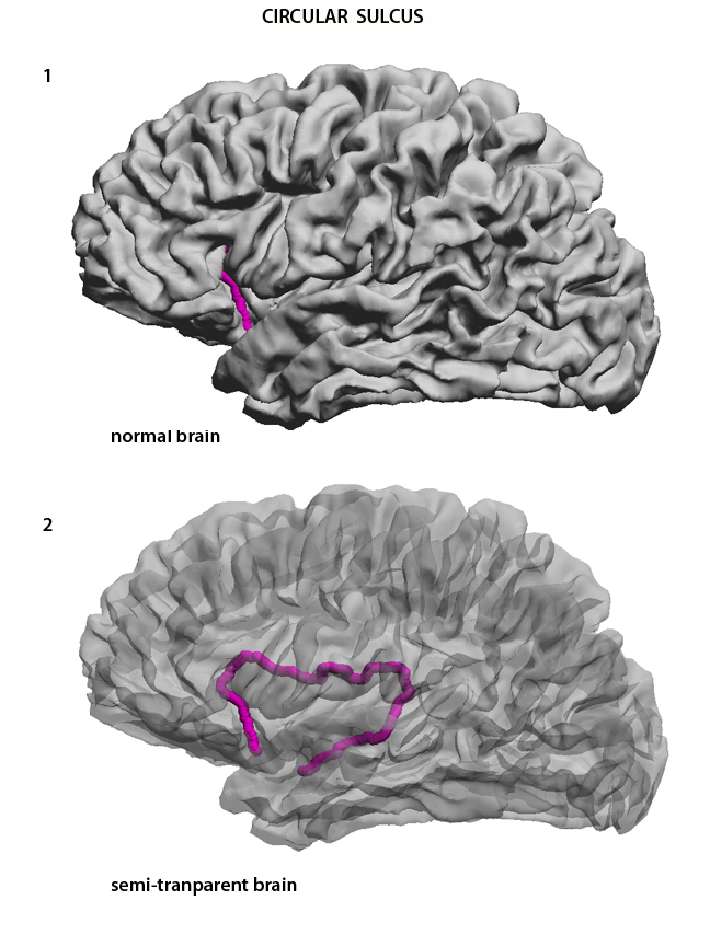
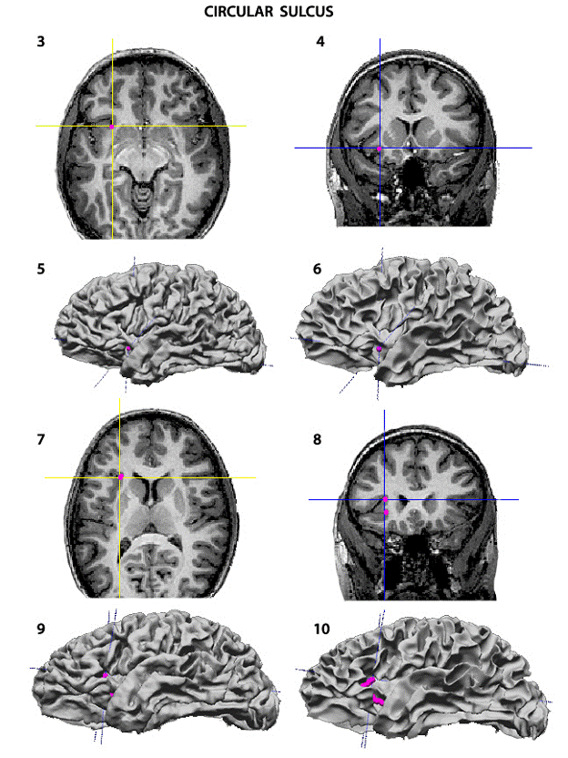
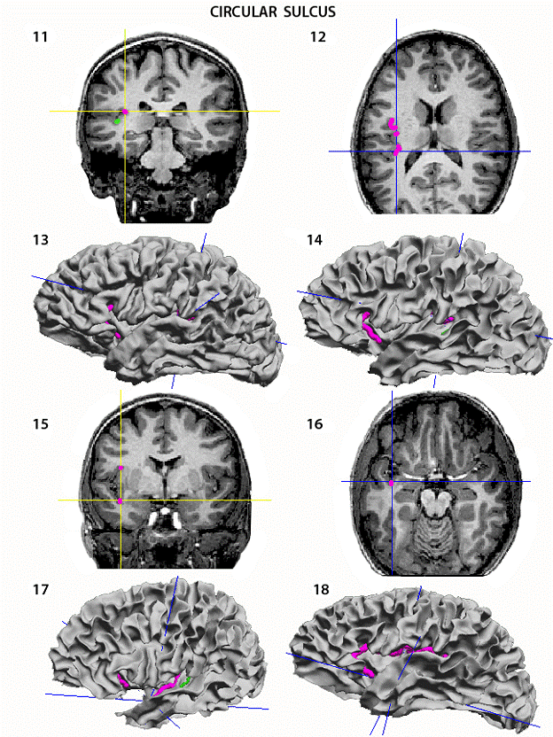
The Circular Sulcus (circS) is the sulcus that runs all around the insula, separating it from the temporal, frontal and parietal lobes. This sulcus cannot be easily followed on the surface rendering because it is always partially covered. To trace it, it is best to use a combination of axial and coronal slices. The first two images show the circular sulcus appearing anteriorly under the frontal operculum (1), and in its entirety in a semitransparent brain (2). Point #1, inferior-anterior: Start with the most anterior and inferior point. This point can be found in axial slices. Go down until the insula and the frontal lobe loose their continuity. Then slowly come up and drop the point at the level at which the insula is first clearly demarcated from the frontal lobe. Drop the anterior starting of the curve (3). In (4) there is the image of this dot on a coronal slice. It can also be seen in the surface rendering. In (5) on the midcortical surface, the one used for the tracing, and in (6) on a gray/white junture rendering. Point #2, superior-anterior: Next move the axial slices up until the insula and the sulcus are not clearly seen any more. Drop down to the highest slice where they can be seen and drop the 2 nd point (7). In (8) the second point is seen in a coronal slice, and in (9) and (10) on the two surface renderings. Point #3, superior-posterior: Use now the coronal slices and move back to the posterior end of the insula. At the point where the transverse temporal gyrus seems to continue into the overlying parietal lobe (TTS in green on the coronal slice) drop the posterior point (11). The axial slice is in (12) but this slice does not provide good reference for the posterior and superior point in the curve. In (13) and (14) are the two surface renderings showing the curve so far drawn. Point #4, end point: In the coronal slices move anteriorly to a few cuts behind the image where the first point appears in coronal slices, so as to clearly see the lowest sector of the insula as it connects to the temporal lobe. This will happen at the beginning of the temporal stem (15). This will be the end point of the circular sulcus. In (16) is the point in the axial slice, and in (17) on the gray/white juncture surface rendering. In (17) the anterior and posterior segments can be seen, in (18) the superior and middle segment is visible.
|
|
USC Biomedical Imaging Research Lab © 2009, designed by Dimitrios Pantazis |