Tutorial 2: Import the subject anatomy
Authors: Francois Tadel, Sylvain Baillet
Download
The dataset we will use for the introduction tutorials is available online.
Go to the Download page of this website, and download the file: sample_auditory.zip
- Unzip it in a folder that is not in any of the Brainstorm folders (program folder or database folder).
- This is really important that you always keep your original data files in a separate folder: the program folder can be deleted when updating the software, and the contents of the database folder is supposed to be manipulated only by the program itself.
Create a new subject
The protocol is currently empty. You need to add a new subject before you can start importing data.
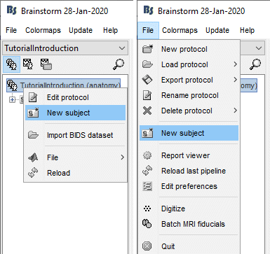
- Switch to the anatomy view (first button just above the database explorer).
Right-click on the top folder TutorialAuditory > New subject.
Alternatively: Use the menu File > New subject.

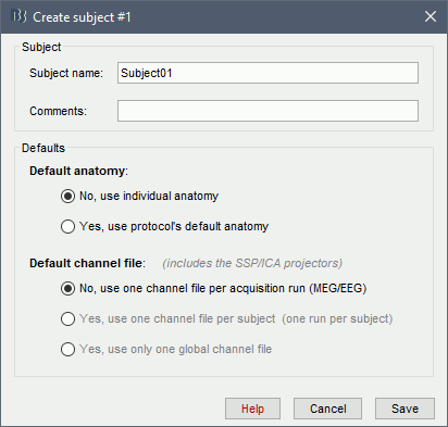
The window that opens lets you edit the subject name and settings. It offers again the same options for the default anatomy and channel file: you can redefine for one subject the default values set at the protocol level if you need to.

- Keep all the default settings and click on [Save].
Import the anatomy
For estimating the brain sources of the MEG/EEG signals, the anatomy of the subject must include at least by three files: an MRI volume, the envelope of the cortex and the surface head of the head.
Brainstorm cannot extract the extract the cortex envelope from the MRI, you have to run this operation with an external program of your choice. The results of the MRI segmentationobtained with the following programs can be imported automatically: FreeSurfer, BrainSuite, BainVISA and CIVET.
The anatomical information of this study was acquired with a 1.5T MRI scanner, the subject had a marker placed on the left cheek. The MRI volume was processed with FreeSurfer 5.3, the result of this automatic segmentation process is available in the downloaded folder sample_auditory/anatomy.
- Make your that you are still in the anatomy view for your protocol.
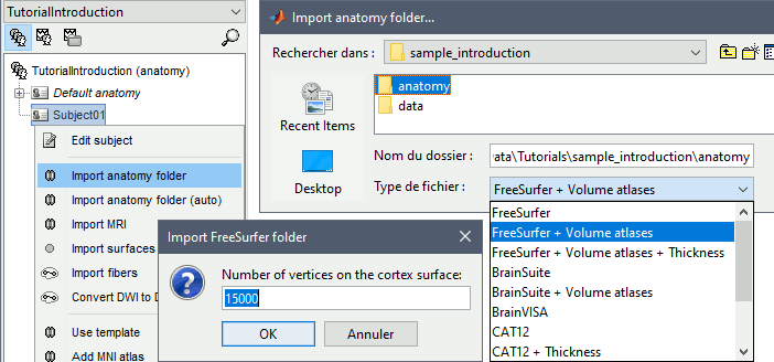
Right-click on the subject folder > Import anatomy folder:
Set the file format: FreeSurfer folder
Select the folder: sample_auditory/anatomy
- Click on [Open]
Number of vertices of the cortex surface: 15000 (default value)
This option defines the number of points that will be used to represent the cortex envelope. It will also be the number of electric dipoles we will use to model the activity of the brain. This default value of 15000 was chosen empirically a good balance between the spatial accuracy of the models and the computation speed. More details later in the tutorials.

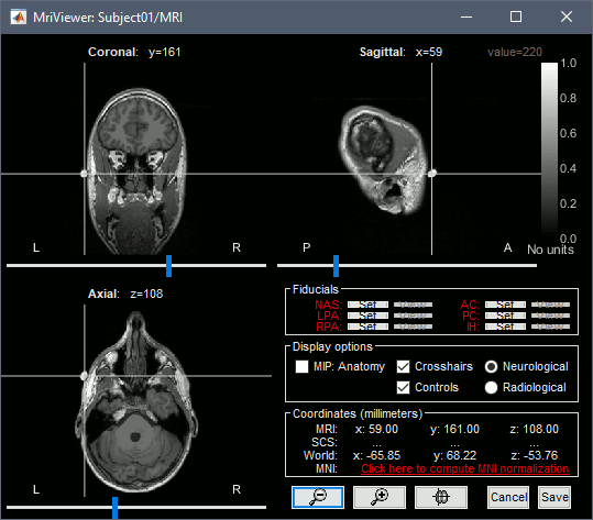
The MRI viewer is displayed, together with a message box that tells you what to do. Follow these instructions. The MRI views should be correct (axial/coronal/sagittal), you just need to make sure that the marker on the cheek is really on the left of the MRI. The you can proceed with the fiducial selection.

Select the reference points
The MRI is already well oriented so you can directly process with the fiducials selection: Nasion, left ear, right ear, anterior commissure, posterior commissure, and inter-hemispheric point. For instructions to find those points, read the following page: CoordinateSystems.