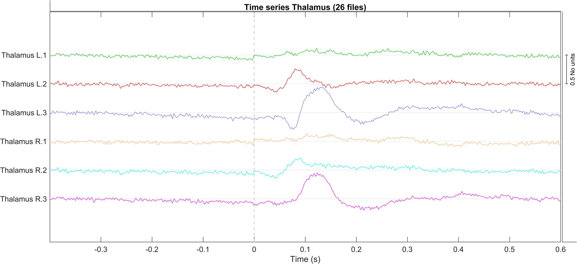
After using the MN imaging with 'dSPM standardisation' and 'unconstarined dipole orientations', the following results are plotted for the scout 'Thalamus'.
I was wondering which directions corresponds to which number. Am I right that L.1 = Left thalamus x-direction, L.2 = Left thalamus y-direction, and L.3 = Left thalamus z-direction?
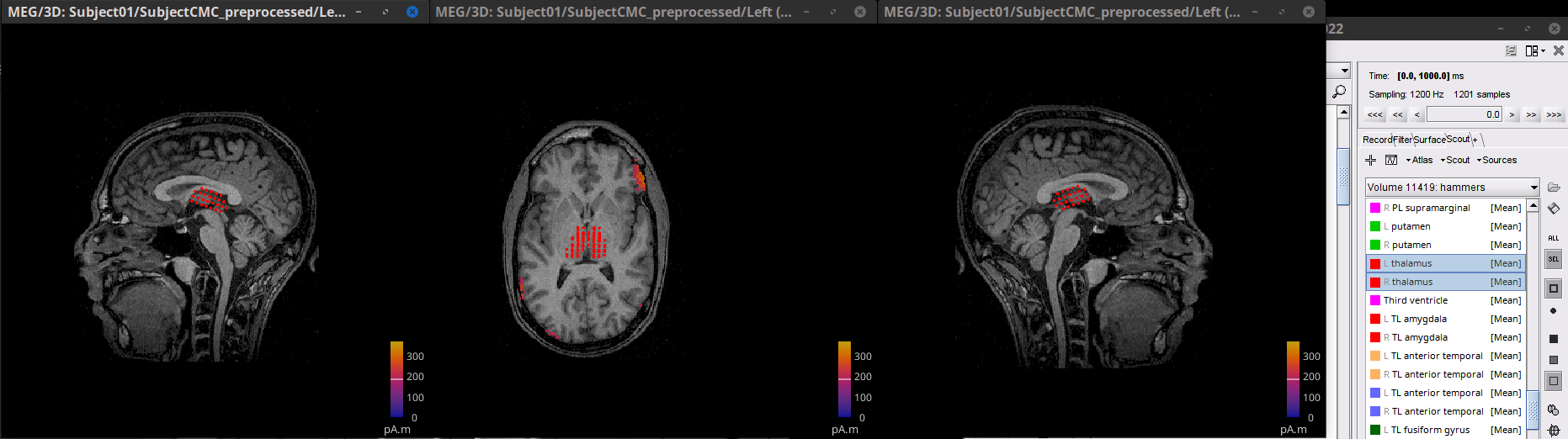
And is it possible to visualize this in the MRI/scout? So what would be the x,y,z direction?
Can you then assume that the activity shown in the figure corresponds to the direction in which it is measured? Since the magnetic field is perpendicular to the electric field.
Sorry to bother you again, so can I say that the activity visible in (seeing the figure from the post above) the L.1 and R.1 (the x-direction) is from the origin to the nasia?
So from the thalamus to the anterior cingulate cortex and the prefrontal cortex?
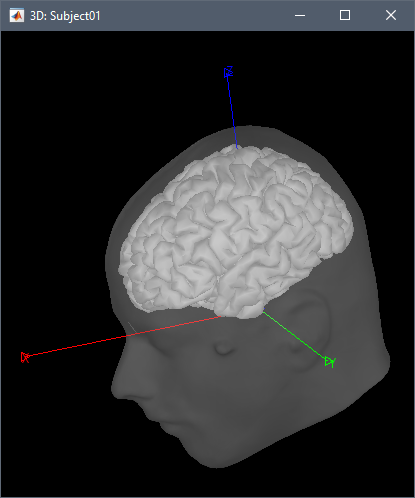
To visualize the axes (X=red, Y=green, Z=blue): open any 3D figure, right-click > Figure > View axis.
L1 and R1 correspond to the amplitude along the red axis.
So from the thalamus to the anterior cingulate cortex and the prefrontal cortex?
Note that this indicates the orientation of the dipoles within a specific brain tissue, it does not indicate that there is current flowing or any form of connection from the cingulate cortex to the prefrontal cortex.