Hello!
I don’t have the anatomical files processed in FreeSurfer, so I uploaded the T1w.nii.gz file of the head before preprocessing to Brainstorm. I then ran FreeSurfer with 10,000 vertices for the cortex surface.
I followed the tutorial https://neuroimage.usc.edu/brainstorm/Tutorials/LabelFreeSurfer to run FreeSurfer from Brainstorm.
During the process, I got a message asking whether to adapt the volume immediately, and I clicked Yes. Everything seems to have gone fine, but among the files created by FreeSurfer, when I try to open ASEG, DKT Desikan-Killiany, and Destrieux, I get an error message saying:
"The dimensions of the two volumes don’t match."
Do you know why this happens? Is it important to fix it immediately? Is there a rigorous way to fix this without rerunning the full FreeSurfer reconstruction, which takes several hours?
Thanks.
Thanks in advance for your help!
What is happening is that FreeSurfer can (and often) change the volume dimensions, voxel size and of the input volume. So, after running FreeSurfer you still need to import to folder with the results. This will replace the original MRI with the T1.mgz MRI in the mri folder within the FreeSurfer results. This T1.mgz has the same volume size as the imported atlases.
As indicated in the tutorial:
[Section: Run FreeSurfer from Brainstorm] At the end, you need to call manually the menu Import anatomy folder described below.
https://neuroimage.usc.edu/brainstorm/Tutorials/LabelFreeSurfer#Import_the_results_in_Brainstorm
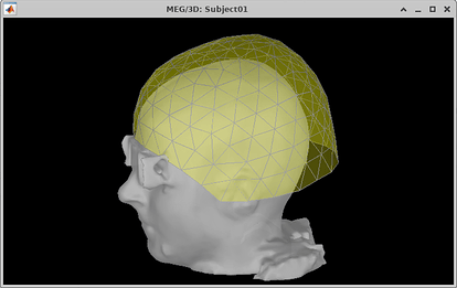
Thanks Raymundo, your suggestion helped me solve the problem. I re-imported the anatomy folder, but now the head mask has a bump in the area of the eyes. Is this caused by the way I imported the data?
Also, since I had already done the MEG data analysis in the functional view do you suggest to do it again? I tried to check the MRI registration, but when I try to refine it, I am told that there were not enough digitized points to perform the refinement.
Thanks for your help.
This is not related to importing the data, but to the MRI data itself. As FreeSurfer results do not have the scalp surface, this one is computed by Brainstorm from the MRI, using default parameters.
Can you share a screenshot of the scalp surface you got?
You can try:
-
Tune-up the parameters to improve the scalp surface.
Right-click on the MRI (once everything is imported), then MRI segmentation > Generate head surface
The default parameters are:
- Number of vertices: 10 000
- Erode factor: 0
- Fill holes factor: 2
- Background threshold: This is guessed from the MRI data
-
Compute the BEM surfaces using FieldTrip method.
Right-click on the MRI (once everything is imported), then MRI segmentation > Generate BEM surfaces > FieldTrip
 Please update your Brainstorm instance to 20-Nov-2025, as there was a bug with the interaction of FieldTrip and SPM plugins, that blocked the computation of BEM surfaces with FieldTrip
That depends on what do you mean with "done the MEG data analysis".
Processing that do not depend on the position of the MEG with respect to the head are ok:
- ICA and SSP
- Frequency filters
- Event detection
Processing that depend on the position of the MEG with respect to the head must be redone:
- MRI-registration (of course)
- Computation of head model, and consequently
- Source estimation and their analysis
Does your data have digitized points for the head shape?
https://neuroimage.usc.edu/brainstorm/Tutorials/ChannelFile#Automatic_registration
 If you have digitized points for the head shape, you need first to compute an accurate head (scalp) surface
Thanks! Attached is the screenshot. I have no digitezed head points.
