I have imported a lesioned brain into Brainstorm after segmenting in FreeSurfer. However, now I have a question about the various views able to be selected in the 3D viewer (by right clicking the image and selecting "views").
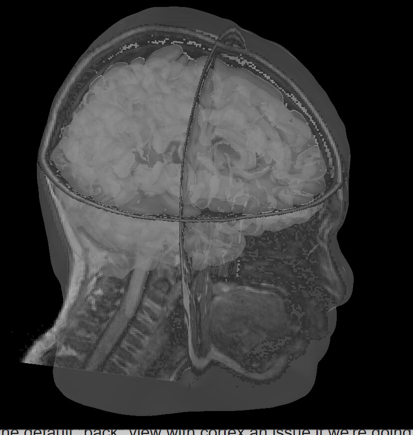
When viewing the reconstructed cortex (lower # of vertices) with the scalp, I click the "view" button, select "back" and get the attached screenshot, which shows that the view is coming from the back but not beginning below the neck.
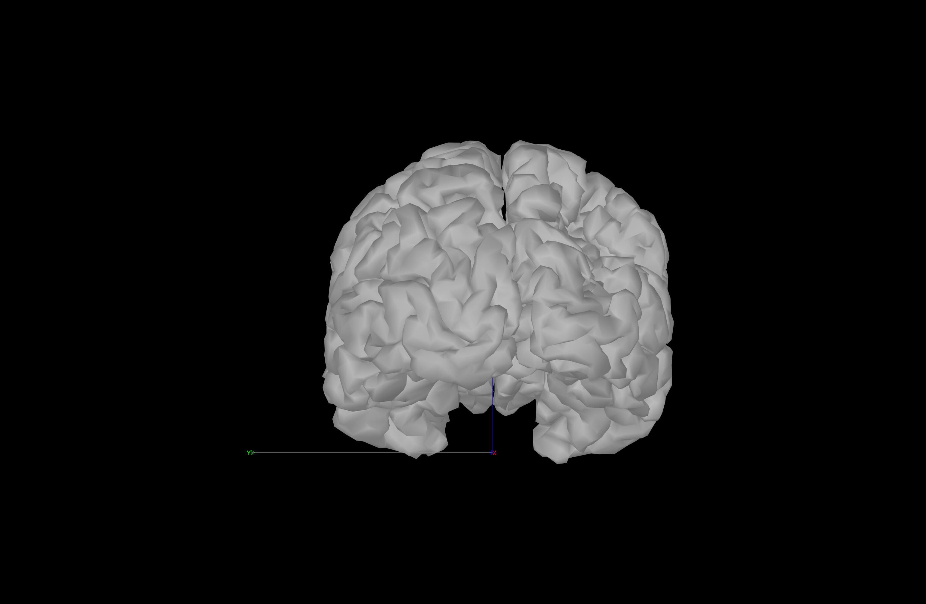
Next, I click the same view (back) with only cortex (again, lower # vertices) and it looks fine. Note that curve at the back of the brain interhemispheric fissure is present in the raw T1w MRI, so this is expected).
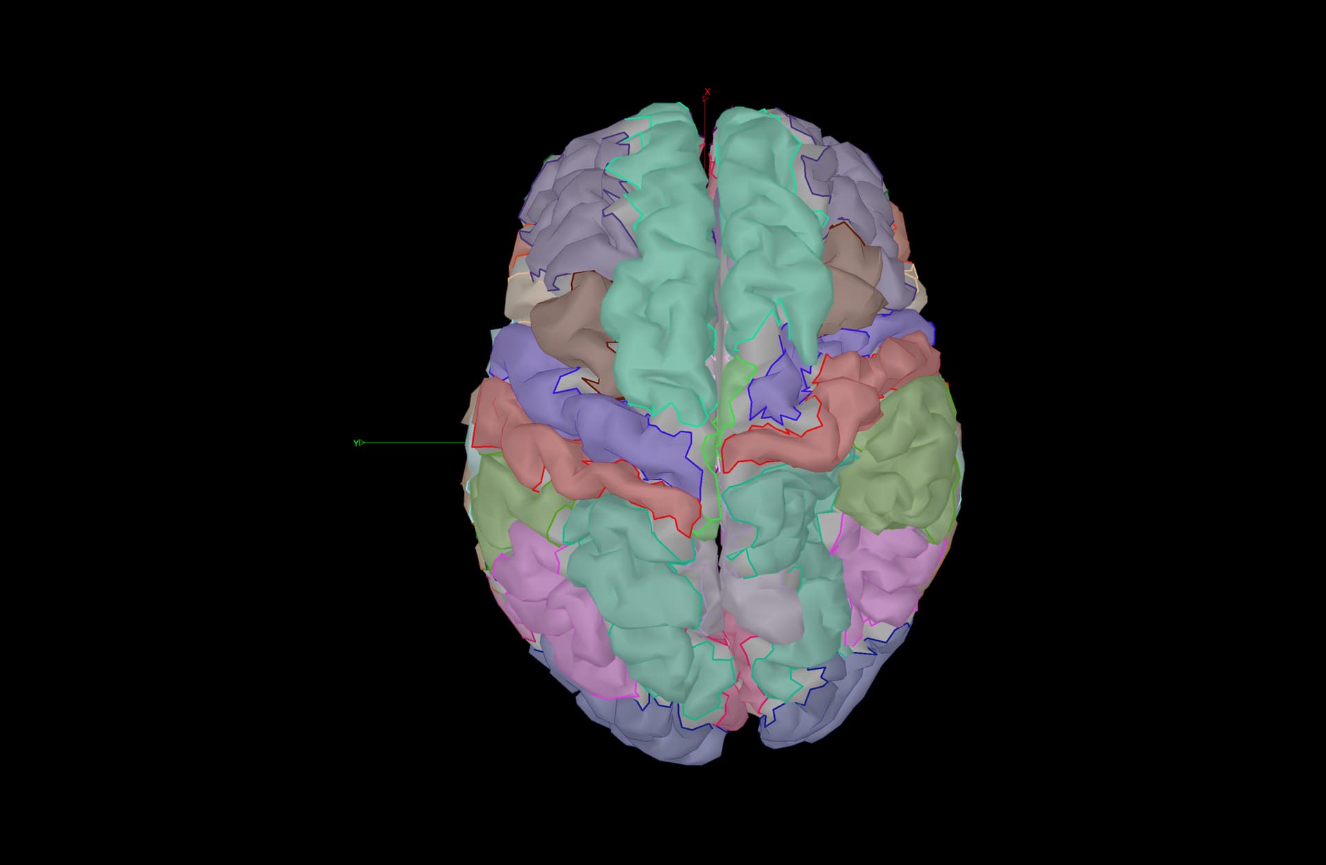
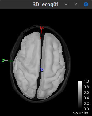
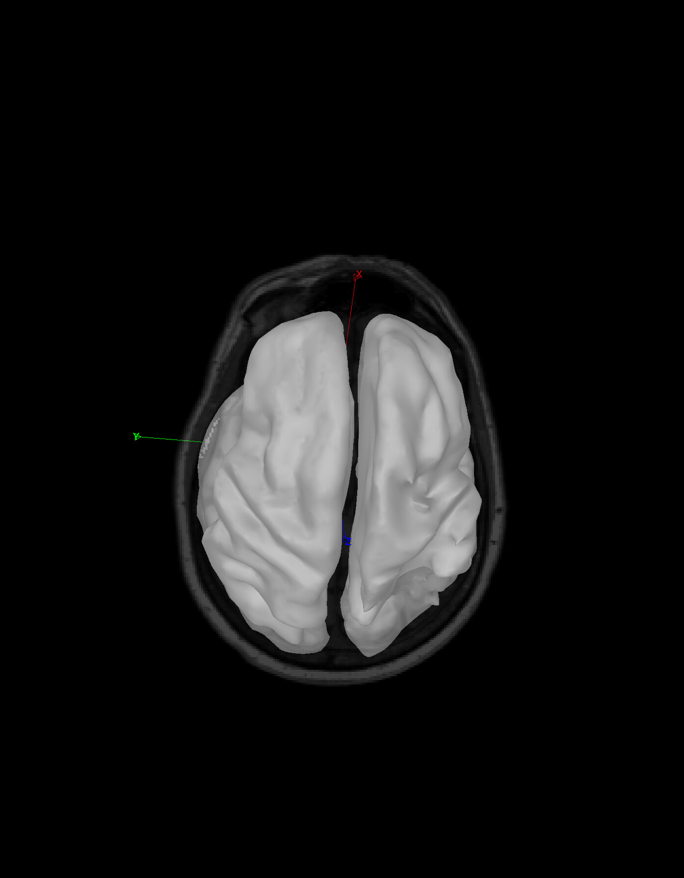
Next, I can view from the top with the Desikan atlas, and segmentation looks ok. Still lower # cortical vertices.
My question: is how Brainstorm registers the default "back" view with cortex an issue if we're doing sEEG source localization? If so, how do we alleviate this?I want to ensure that FreeSurfer/Brainstorm has registered our brains correctly so we may correctly do our analyses. Please let me know any thoughts. Thank you very much!!
But the alignment of the head and the brain of the same subject will be in the same coordinate system and with have the same orientation.
Brainstorm use different approaches to estimate the scalp surface (from a simple to a more realistic shape). Still, all of them are based on the orientation of the cortex, which is obtained from any segmentation tools and then reorientation to the SCS coordinate system when you load the MRI or compute the MNI coordinates.
(https://neuroimage.usc.edu/brainstorm/CoordinateSystems)
SCS is based on: Nasion, left pre-auricular point (LPA), and right pre-auricular point (RPA).
Origin: Midway on the line joining LPA and RPA.
Axis X: From the origin towards the Nasion (exactly through).
Axis Y: From the origin towards LPA in the plane defined by (NAS,LPA,RPA), and orthogonal. to X axis.
Axiz Z: From the origin towards the top of the head.
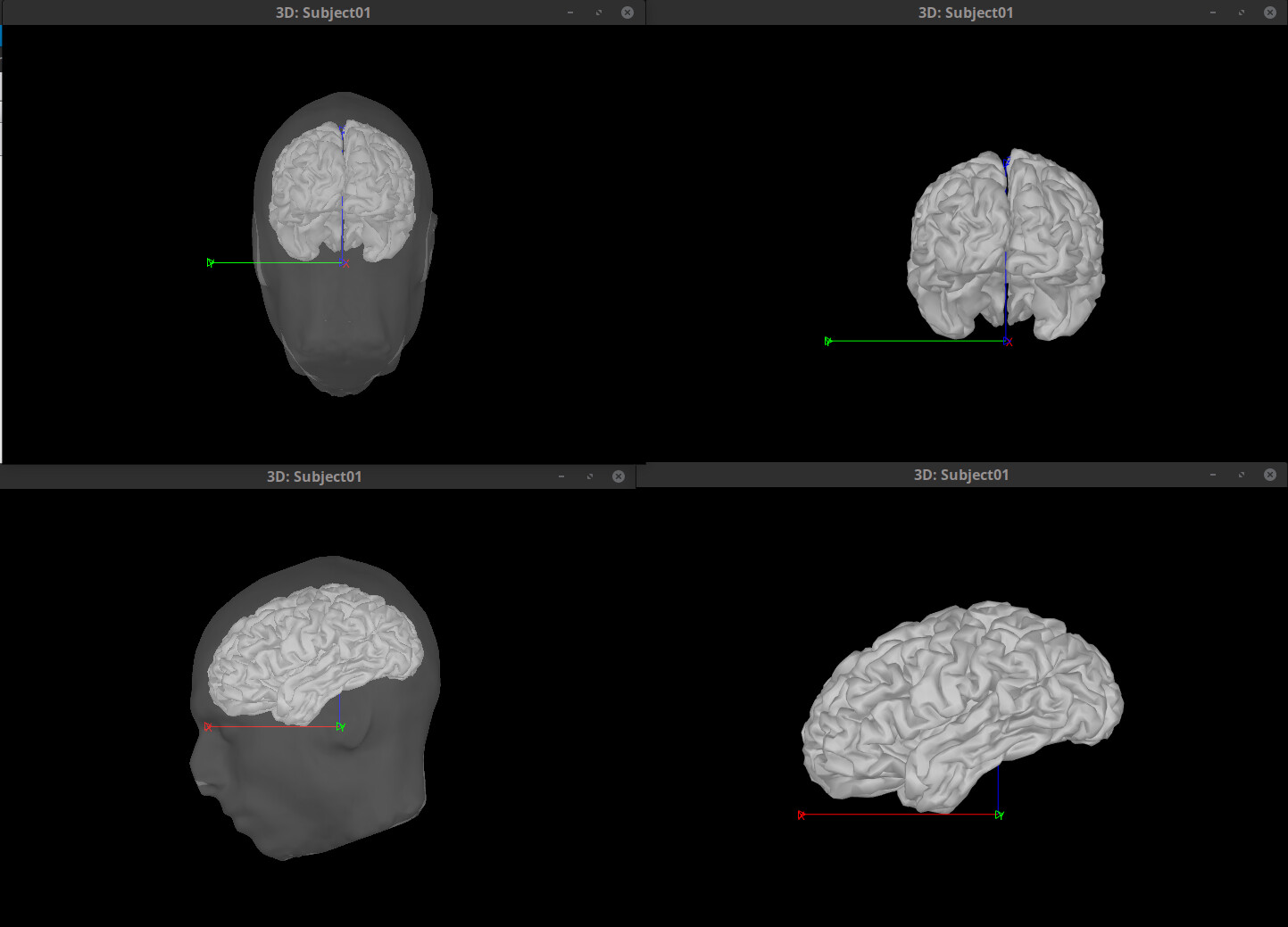
In the figures, you can show the SCS axes with right-click > Figure > View axis (only the positive side of the axis is shown). X:red, Y:green, Z:blue. As such, the Back view corresponds to the view from a point from the -X axis (top figures); the Left view is the view from +Y axis (bottom figures), etc.
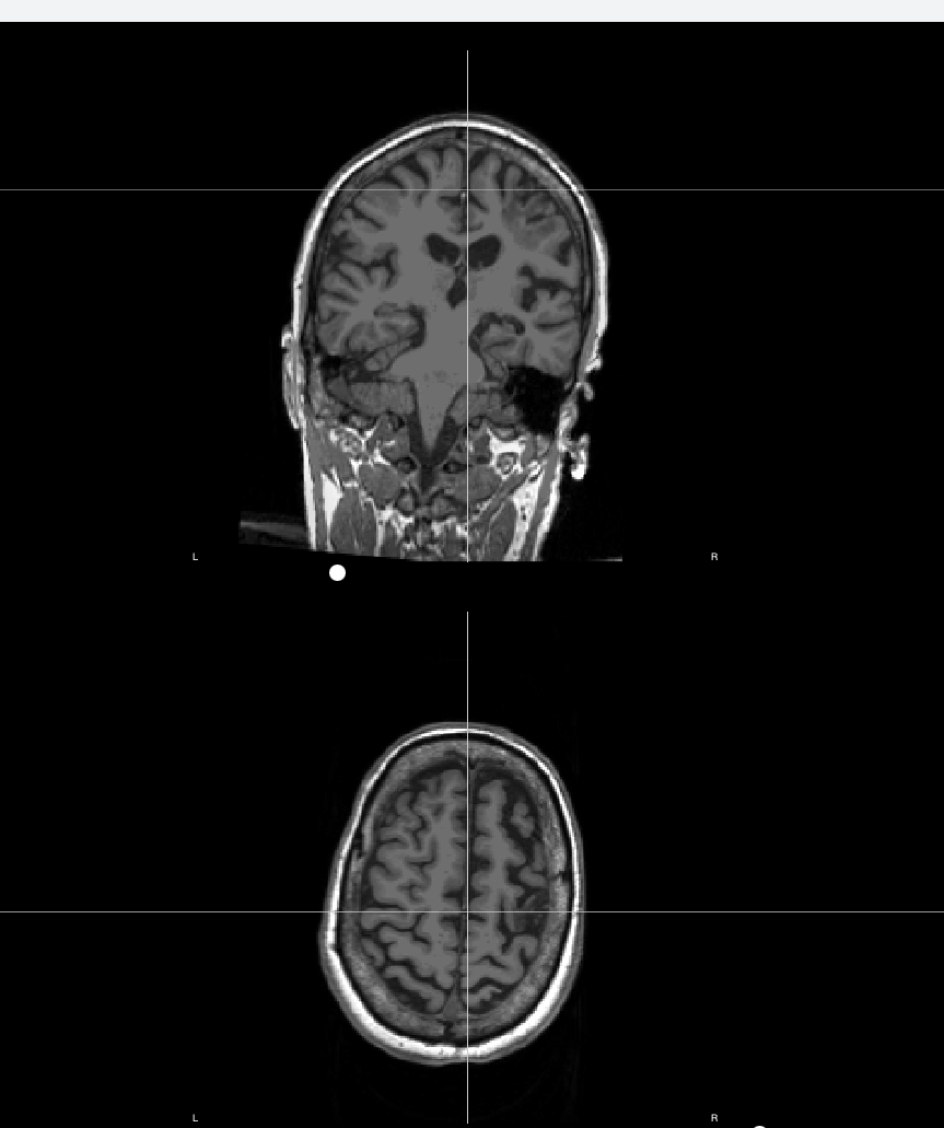
I'm wondering about how a crooked interhemispheric (IH) fissure affects the registration. As you'll see below, there is a crooked IH fissure and it seems that the crosshairs do not align with the IH fissure regardless of the height/depth of the crosshairs along the fissure. What do you recommend for managing this? Another consideration is that the optimal respective slices for the AC/PC points for this scan are 5 or 6 slices away from each other laterally. Perhaps this corrects for the crooked IH fissure? I'm not sure. Just want to give you the most information.
This happens as the axes in SCS (coordinate system used in Brainstorm) are defined with the Naison and pre-auricular points. There is nothing to worry here, as you can see in all the figures the cortical surface and the anatomy are properly registered.
A clarification on this point:
IF MRI displayed in the figure (as it is the case in your figures), the views use the orientation of the slices, instead of the orientation of the SCS axes.
Thank you. I want to clarify a point about the crosshairs & IH fissure.
The screenshot I included above with the crosshairs & IH fissure is upon import before the nasion & peri-auricular points are defined, which leads me to believe that this person's head was tilted in the scanner, perhaps?
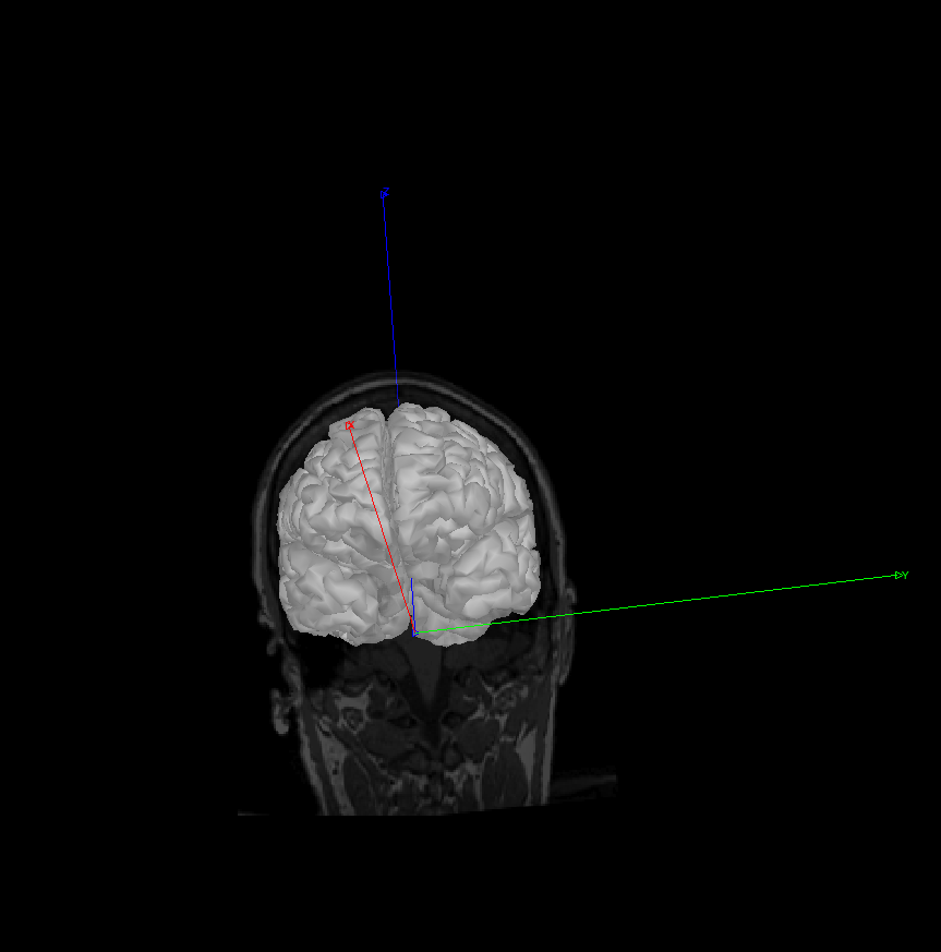
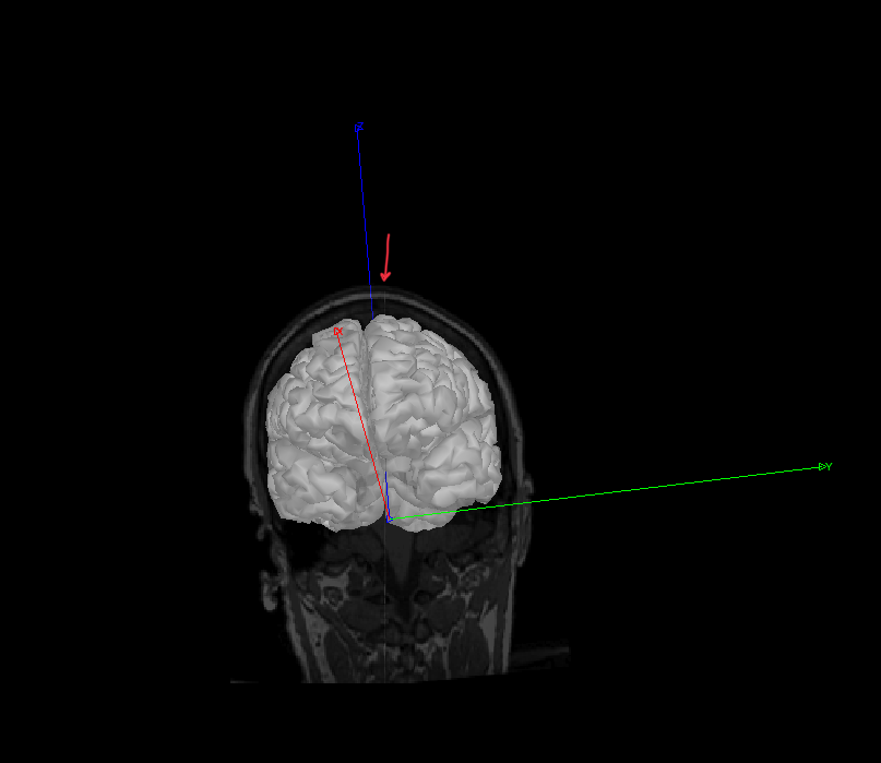
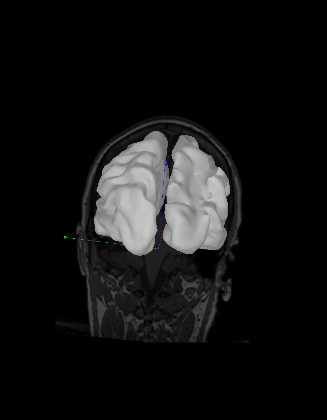
Regardless, here are a couple more screenshots of this person's registration after defining the nasion, LPA/RPA, AC/PC. I'm wondering why the midline of their skull/head (I attempted to denote this with my red arrow) is not aligning with the IH fissure. What do you think of these screenshots?
Note that I slightly rotated the image along the x axis so that you could just see the midline of the skull.
Please let me know if you need more info. Thanks again.
It seems the IH fissure is aligned with the X (red) axis. However, as the MRI volume is tilted, it gives a impression IH and the red axis are not align.
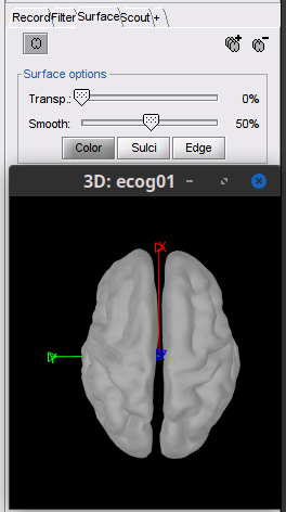
Try this approach to verify the alignment.
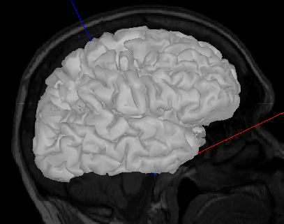
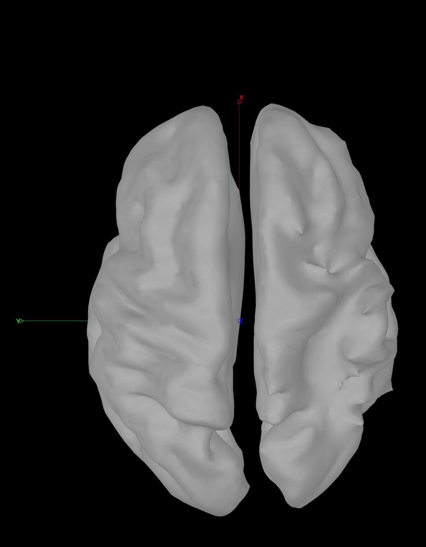
Plot only the cortex (not the MRI slices), show the axis, smooth the surface (50%), change the view to top. The red axis should run parallel to the IH fissure.
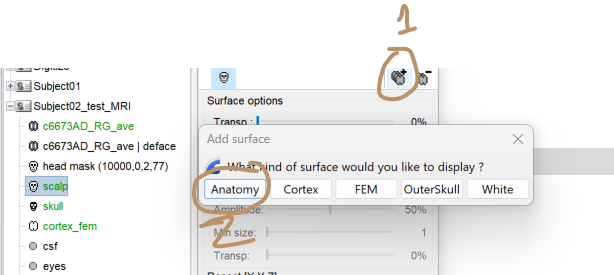
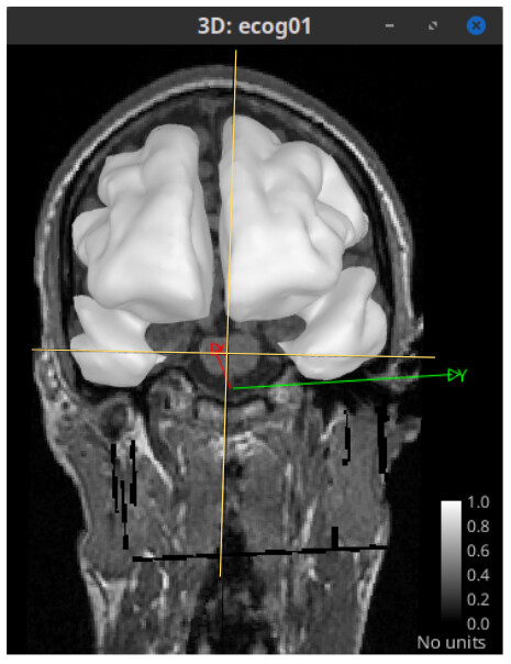
Now, add the MRI slices. In the Surface tab, click on "Add a surface" (), and add the Anatomy layer.
Note that the MRI slices are not aligned. The yellow lines show the slices planes:
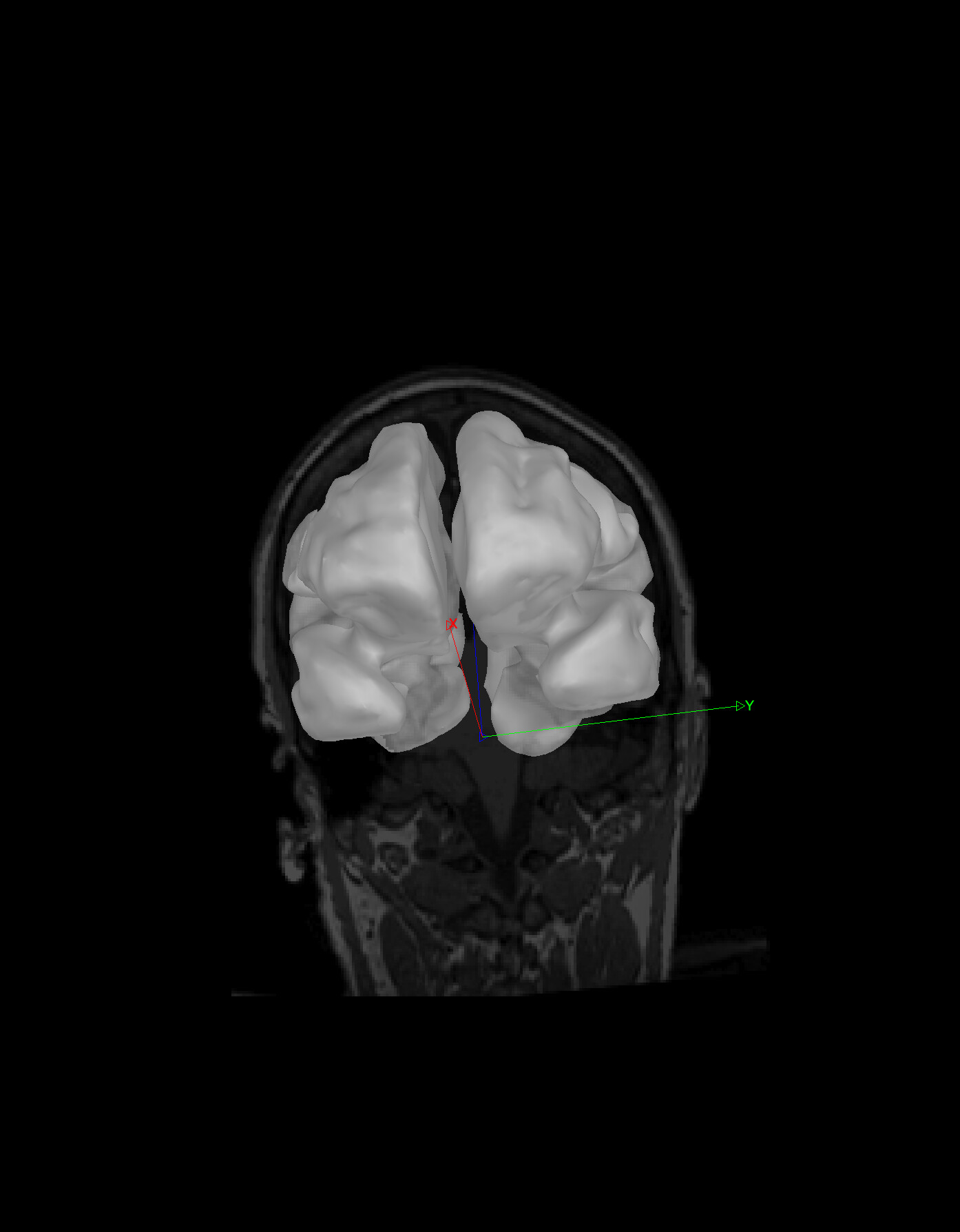
Thank you for your reply. I did what you said by first adding the cortex smoothed 50%, then adding the anatomy over top of cortex. Below are my results; in short, you seem to be correct about the red X axis aligning with the IH fissure. I didn't know how to get the yellow crosshairs that you have in your third figure.
There are screenshots at the bottom of this post.
My questions are:
1 If this needs to be adjusted, how should I go about making these adjustments, and how can I prevent misalignment from occurring in the future?
2. Is this misalignment related to where the fiducial points are? Note: our patients have had a stroke, so there are stroke lesions to the brain.
3. How would this misalignment in the current form impact EEG source localization?
My bad, I added those yellow lines on the screenshot I took to illustrate where the sagittal and axial planes were. A way to see where are these slice planes is to plot the MRI in 3D view, then set to front (or back) view and slighly change the point-of-view to see the border of the sagittal and axial slices.
Thank you for the explanation.
Could you please clarify what you mean by preprocessing the MRI volume? I ran the recon-all script of FreeSurfer on our participants' MRI scans. Is this what you meant?
Also, just to confirm: you're saying there's nothing to adjust with this scan even though the midline of the head is "off" from the IH fissure? And that everything looks as it should.