Hi @edelaire, that's a interesting question.
Here my comments on the post.
I'm also pinging @John_Mosher, who can certainly give you more details on the topics I am not replying to.
The should figure is not the dipoles, but the leadfields for that sensor (Fp1). For the blue arrows, the orientation will show orientation of the dipole in that direction that will have the maximum influence in the given sensor. While the magnitude shows the sensitivity of the given sensor to that (vertex) location. You can compare the leadfield vectors for EEG and MEG, the EEG ones are radial, while the ones for MEG are tangential.
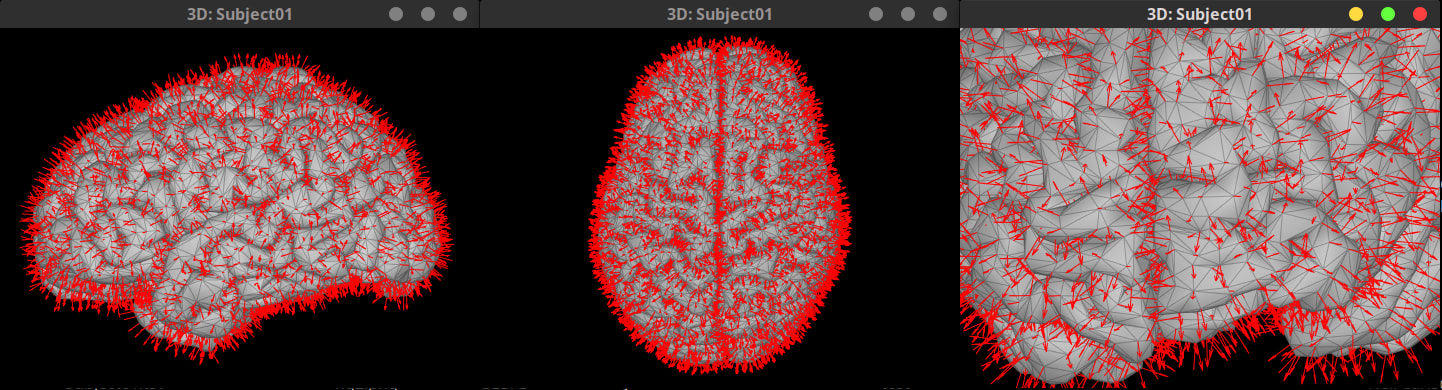
The normals for the vertices are stored in the surface file. AFAIK there is not a way to plot them from the GUI, but it can be easily done.
- Plot the cortical surface
- Export that cortical surface to Matlab as variable, e.g.,
surfo - Plot the normals with this snippet:
hold on
quiver3( ...
    surfo.Vertices(:,1), surfo.Vertices(:,2), surfo.Vertices(:,3), ...
    surfo.VertNormals(:,1), surfo.VertNormals(:,2), surfo.VertNormals(:,3), 3, 'r' ); % Scale by 3, for visualization
That's correct, it's an interpolation for visualization. If it were done face wise, the maps would be piecewise, and that would not really present the data on the cortex that is assumed to be continuous.
Correct, just for visualization. Since the dipole currents are located in the vertices, Scout aggregations are properly handled as the Scouts are sets of vertices.
One potential solution for this visualization issue, that we have not implemented, is to draw the Scouts using the midpoint in the mesh edges.
The normal to the vertex is the average of the normals of the adjacent faces. See tess_normals.m. This measure has smoother transitions (following the assumption of continuos surface) than the face normals which change abruptly between adjacent faces.
Similar to the normals, the areas in the faces can vary a lot for adjacent faces. The area that represents each vertex dipole is set to sum of 1/3 or the adjacent face areas. In this sense, the vertex areas have less variations as it is balanced by the different adjacent face areas.
Have you encounter this post?
In the pragmatic aspect, using vertices as dipoles simplifies the equivalence between the anatomy and the headmodels, this helps to the use of cortical atlases which are defined vertex-wise. Most mesh operations are computing on vertices rather than faces.
I'll let @John_Mosher and @tmedani to provide further comments on this.