Thank you for the detailed response. I will try to keep it simple and edit channel file and MRI registratoin on the channel file, rather than a separate SEEG/ECOG registration then adding positions to channel file.
5 more questions as follow-up:
- I changed channel group as SEEG for all electrodes and I renamed all the electrode groups by letters ascending alphabetical order, but when I go to edit electrodes there is only 1 electrode letter E.
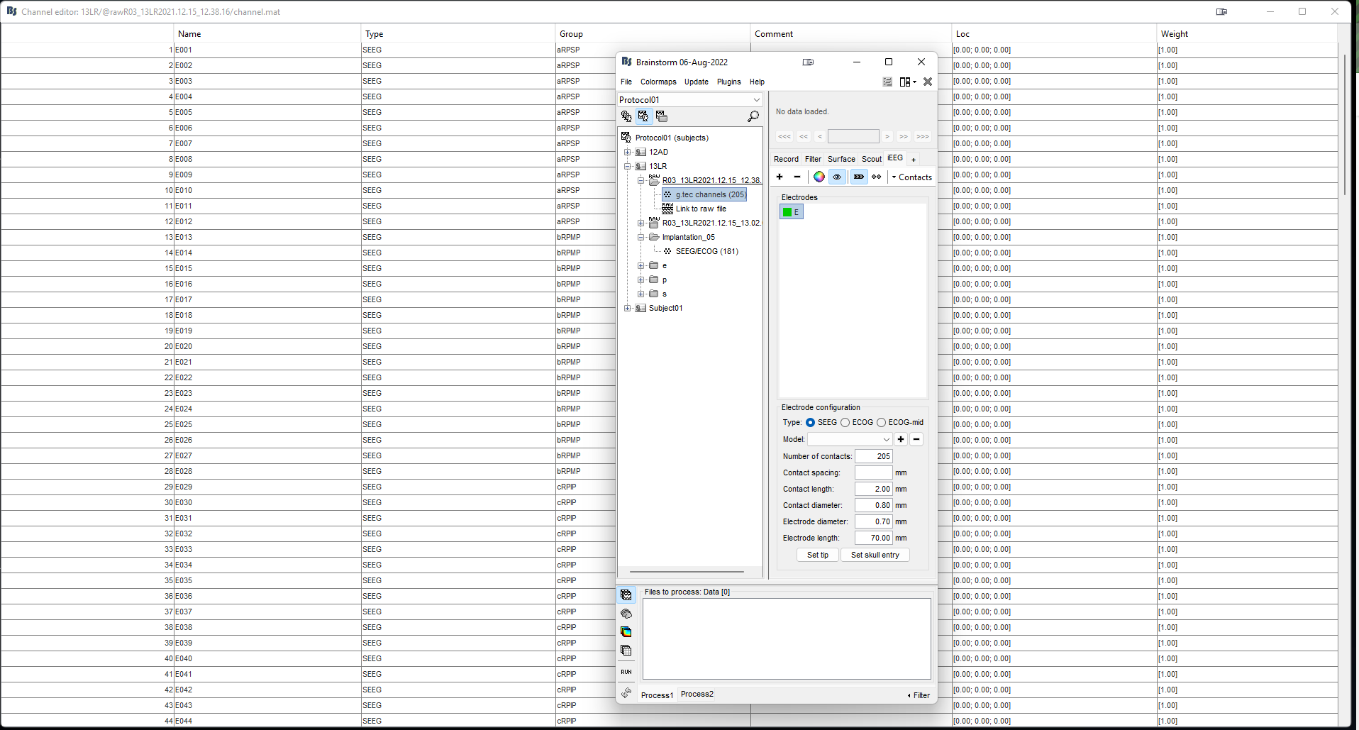
It appears I must change the name of the electrode to different letters for different electrodes.
You see in the screenshot, the electrodes are named ascending numeric order up to total number electrode contacts. I think I'm missing the point of the channel group. I can make groups by lobe or region I guess, but not sure how to easily focus on them.
-
I'm using postop CT SPM reslice for MRI registration to set the electrode tip and entry points. Is it better to use MNI normalized SPM of postop CT?
-
Is Cat12 or Brainsuite preferred for surface generation? Brainsuite takes longer and my computer overheated during processing, CAT12 is faster but the brain looks a little like knife-blade gyri of FTD even though patient only has moderate age-related atrophy.
-
Is iEEG atlas labeling of contacts meant to "find" what tissue space the electrode is going through, gray matter vs white and also if a specific location based on parcellations?
Besides the table format of the information, how else can it be visualized? Is there a way to analyze all the contacts that are in gray matter as a group for example? Or all that are in frontal lobe? -
Any suggestion for the workflow since there are about 15 patients that I'm interested to analyze and each has about 10-16 electrodes totaling 200+ contacts and 5 recording sessions that are about 5 min each. If I edit channel file for one recording of 5min duration and there are 4 other recordings, can I copy the electrode positions and names from the first modified file to the others to save time instead of manually entering letters and repeating the registration/target/entry of electrodes? Moreover there are events that are marked in succession as a single timepoint at time at start of event and then the event ends when the next event starts, but the block of time is not marked from what I can tell.
In the future I'm considering figuring out how to use mia to do group analysis of the different subjects recordings for trends of effects on SEEG signals from the events the amplifier generates/records. Seems like it may influence the answer to above questions.
Many thanks.