Thanks for all the previous help! I really learned a lot from here, and very appreciate this site.
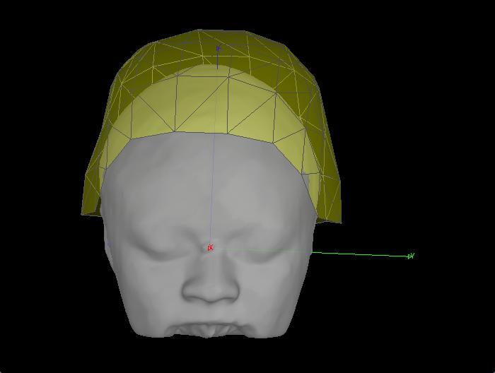
I am trying the polhemus .pos file, and Vitamin E gel at the fiducial points for better co-registration for MEG and individual MRI. Co-registration figures are below:
It is the figure that with both vitamin E gel on fiducial points, and the head shape digitization from Polhemus.
It look like that we have better co-registration with just vitamin E. Is there a way to evaluate how good it the co-registration? What I did is to save the .pos file inside the MEG folder.
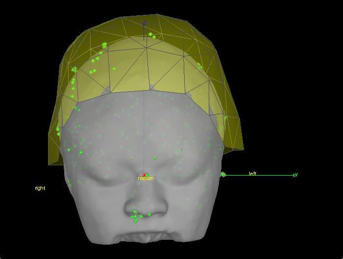
Your headshape points do not look very precise and not regularly spaced. For example, this cloud of points at the tip of the nose is a bit strange. You should try to pick points that are more homogeneously spaced.
Have you used Brainstorm for recording those points? http://neuroimage.usc.edu/brainstorm/Tutorials/TutDigitize
Why do you say that registration #1 is better than #2?
The only way to decide which one is better would be to compare it with pictures of the subject in the MEG…
If you are not happy with the automatic registration fitting, you can skip it, or refine it manually (right-click on channel file > MRI registration > Edit).
About the vitamin E, it probably doesn’t help much for localizing the fiducials. If you use the Brainstorm digitizer, you have the option to pick three anatomical points that are precise and easy to find enough without any additional marker. A marker on one cheek is a good way to identify the left and the right of the T1 scan. http://neuroimage.usc.edu/brainstorm/CoordinateSystems
Thank you so much for the reply.
Thank you for the link for digitization, I will try do the head shape points without drawing continuous lines next time, that’s a better idea. I did the digitization and created .pos file from CTF software, trying to draw lines along the head. This study participant is an African American lady, she has lots and lots of hair, so it is hard to draw lines. I tried to just draw short lines. Probably that makes the points not evenly spaced.
About the vitamin E gel: We are using CTF MEG system, there are three head localization coils, nasion, LPA, and RPA, fixed by donut tape. We left the donut tape on after MEG, and put vitamin E gel on exactly the same spot for MRI. The vitamin E gel could be seen in free surfer processed MRI image. When I import the MEG recordings, the helmet looks in a great position, as in the first image. However, the second image for head shape is by automatic registration fitting (when the .pos file inside the MEG data folder imported), the helmet looks have more overlap with the head surface… I am not sure if that is a way to judge the fitting or not. I wish there is an objective way. I will try the manually refine, thank you so much for the link.
Please feel free to suggest, we are still exploring better co-registration, for sourcing analysis. Shall we take a picture of study participants while they were in the MEG recording position? Comments will be really appreciated.
If you use Brainstorm to get the Polhemus points, you’ll be able to digitize both the position of the CTF coils and the anatomical points.
This is the way we do it at the MNI, and we designed it for a CTF system.
If you simply copy the .pos file in the .ds folder, it should all work nicely.
Hi Xianghong,
Using the vitamin E markers is an excellent way to know where the coils are. This will help you correctly identify the location of the coils on the MRI. You can use only this as a means of co-registration, if desired. But then you should not use the Brainstorm .pos file.
Many sites do not have this option since MRI is not always collected directly after the MEG. Therefore Brainstorm offers an alternative which allows you to collect the positions of the coil AND some well-defined anatomical locations. Then you can also collect a headshape to use for further refining this coregistration. Brainstorm transforms the data to the anatomical locations and therefore you should mark on the MRI the anatomical points - unlike with the vitamin E, you would want to mark on the MRI the coil locations.
I suspect the two methods are not producing the same result because you are mixing them or because your head points appear to be a bit noisy. If you want to collect headshape, we suggest you collect an even distribution of points from the forehead to the inion and down to the ears. A few points on the nose should also be collected.
Please let me know if this is less than clear.
Beth
[QUOTE=ebock;9032]Hi Xianghong,
Using the vitamin E markers is an excellent way to know where the coils are. This will help you correctly identify the location of the coils on the MRI. You can use only this as a means of co-registration, if desired. But then you should not use the Brainstorm .pos file.
Many sites do not have this option since MRI is not always collected directly after the MEG. Therefore Brainstorm offers an alternative which allows you to collect the positions of the coil AND some well-defined anatomical locations. Then you can also collect a headshape to use for further refining this coregistration. Brainstorm transforms the data to the anatomical locations and therefore you should mark on the MRI the anatomical points - unlike with the vitamin E, you would want to mark on the MRI the coil locations.
I suspect the two methods are not producing the same result because you are mixing them or because your head points appear to be a bit noisy. If you want to collect headshape, we suggest you collect an even distribution of points from the forehead to the inion and down to the ears. A few points on the nose should also be collected.
Please let me know if this is less than clear.
Beth[/QUOTE]
Hi Beth and Xianghong,
Glad to see your discussion. I am doing MEG with CTF system recently and I got some questions about co-registration with MNE. We also used vitamin E gel marking the coils position when scan T1 image. It’s very tricky to align the coils position(from MEG functional data) to the marker (VE). It seems that the coils has different orientation and it’s very difficult to make the three digitizer point in the right position meantime. I noticed in brainstorm the digitizer point has no orientation. I wonder whether there should were different orientation for the coils. I was instructed to make sure the lines of the LAR and RAR coils being vertical relative to the ground and nasion coil being horizontal to the ground. But actually I do not know why… I also wonder whether there is an evaluation for the effect of co-registration between the VE marker and the digitizer after what Xianghong has done.