Some of our study participants have CapTrak data, some don't. We measure EEG at three time points (Pre, Post, Follow up). Some have MRI, others don't. This presents two scenarios and a couple of questions:
Scenario 1: Lets say we have CapTrak data for someone for only for 1/3 sessions (with an MRI). Question 1: would it be better to apply these CapTrak channel locations from one session to the other two sessions or instead use the standard electrode positions with manual adjustments to better fit the MRI?
Scenario 2: if we have CapTrak data for someone without MRI (standard ICBM152 2023b anatomy). Question 2: the person's head shape/size may be different than standard anatomy, meaning that when the CapTrak-digitized electrodes are registered to standard MRI, the electrodes may be inside or far away from the standard head.
Since we digitized the electrode positions on the participant's head, we know the location of the channels on their head, suggesting to me that these positions shouldn't be altered other than projecting the electrodes to the surface (to correct for any head size/shape differences). Do you agree with this logic?
Scenario 1: Assuming you're using an electrode cap, I believe the digitized positions would be much more accurate than the standard positions, even if there is a small shift between sessions.
Marc,
Yes, we are using 96-channel ActiCap. Your suggestion makes sense to me that I should use another session's electrode locations from this participant before I used standard electrode layout. Therefore for this person, I will apply one session's CapTrak electrode positions to another session's (the one that had bad CapTrak data).
Raymundo,
I will look into how to warp default anatomy. I was unfamiliar with this technique. I will get back to you about the results I obtain.
Is it necessary to generate a new head model for the session that is using another session's electrode positions? Would it be an identical head model, or is it better to generate a new one? I tried looking through the tutorial but did not find the answer.
My assumption was that I needed to generate to separate head models, but I wanted to confirm.
The head model depends only on the Anatomy and the Channel file (sensors and their location with respect to the anatomy). Thus, it is possible to copy the head model for the sessions using the same channel for the same subject, option Copy to other folders within the right-click menu of the head model.
I followed the tutorial for warping (https://neuroimage.usc.edu/brainstorm/Tutorials/TutWarping#Edit_the_initial_MRI.2FMEG_registration).
First question: is the first paragraph (and the steps prior to the actual warping section) relevant for sEEG, or is this just for MEG helmet with Polhemus digitizer? I see head points on this standard anatomy, but I'm not sure if they're somehow taken from CapTrak data or they are from the standard brain. If from standard brain, I should not edit them and can proceed with warping?
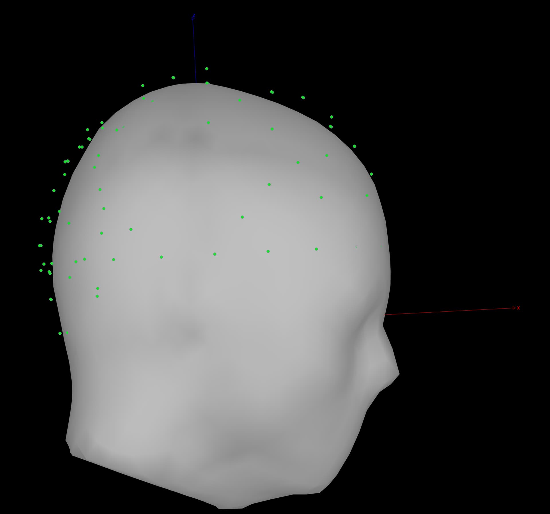
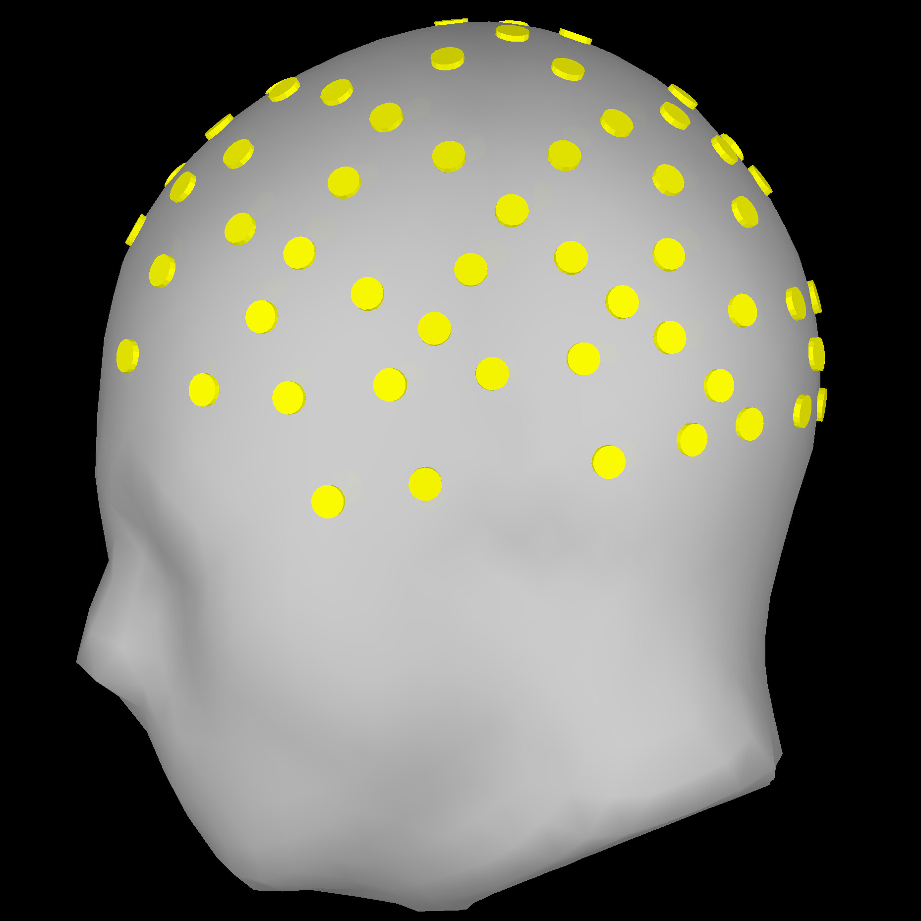
These are the head points I'm referring to (unwarped standard brain):
They seem to look better in the back, but I'm wondering if, assuming I did not need to do anything with the head points (question above), is it okay from here to project electrodes to surface and move forward with processing?
Do you mean the section ** Prepare the anatomy template**?
This step is done to ensure the best alignment between the Default anatomy and the digitized fiducial points. If you use the same fiducial definition, nothing else is needed. Before proceeding with the warping, check the alignment between fiducial sets: right-click on the channel file > MRI registration > Check. If improvement is needed, select Edit.
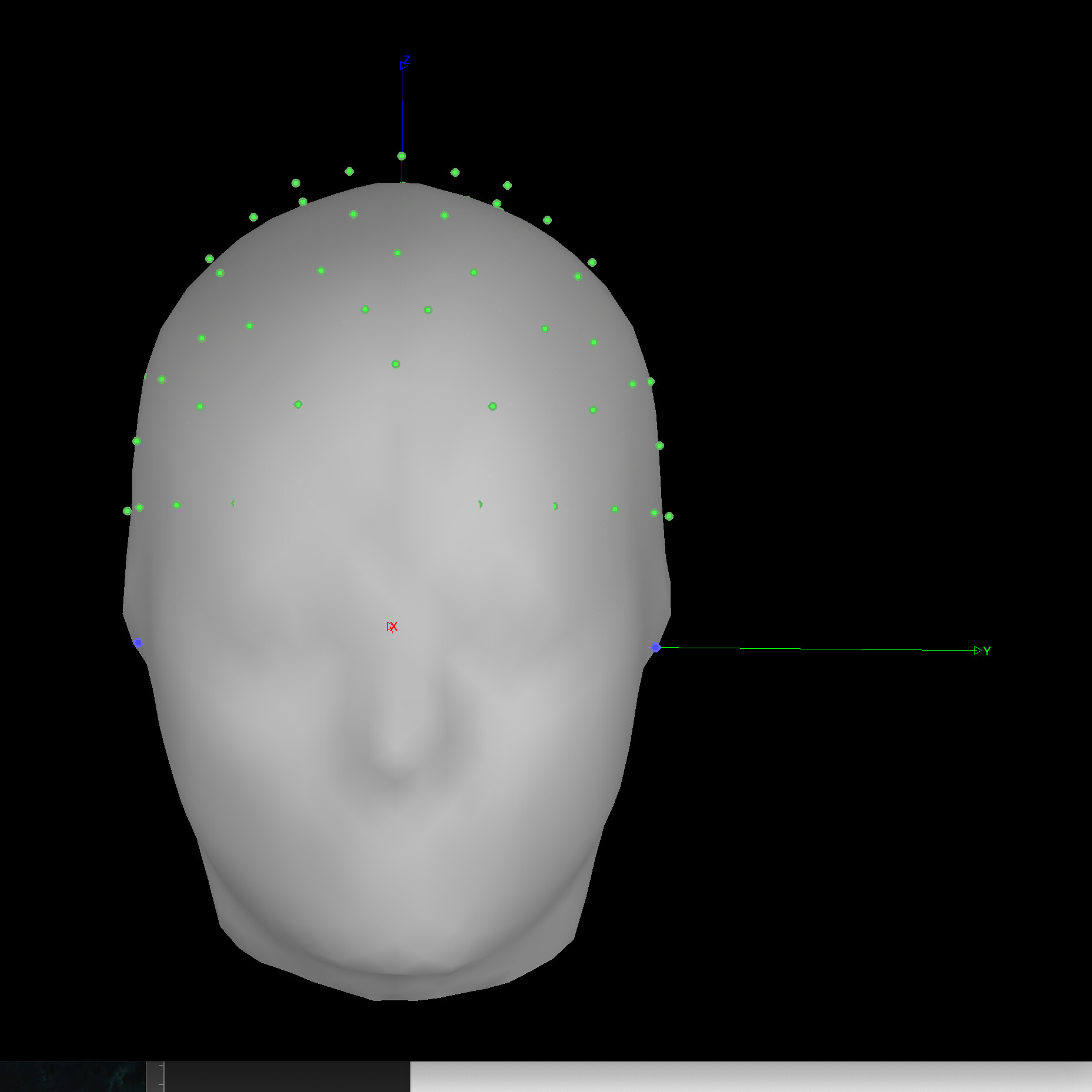
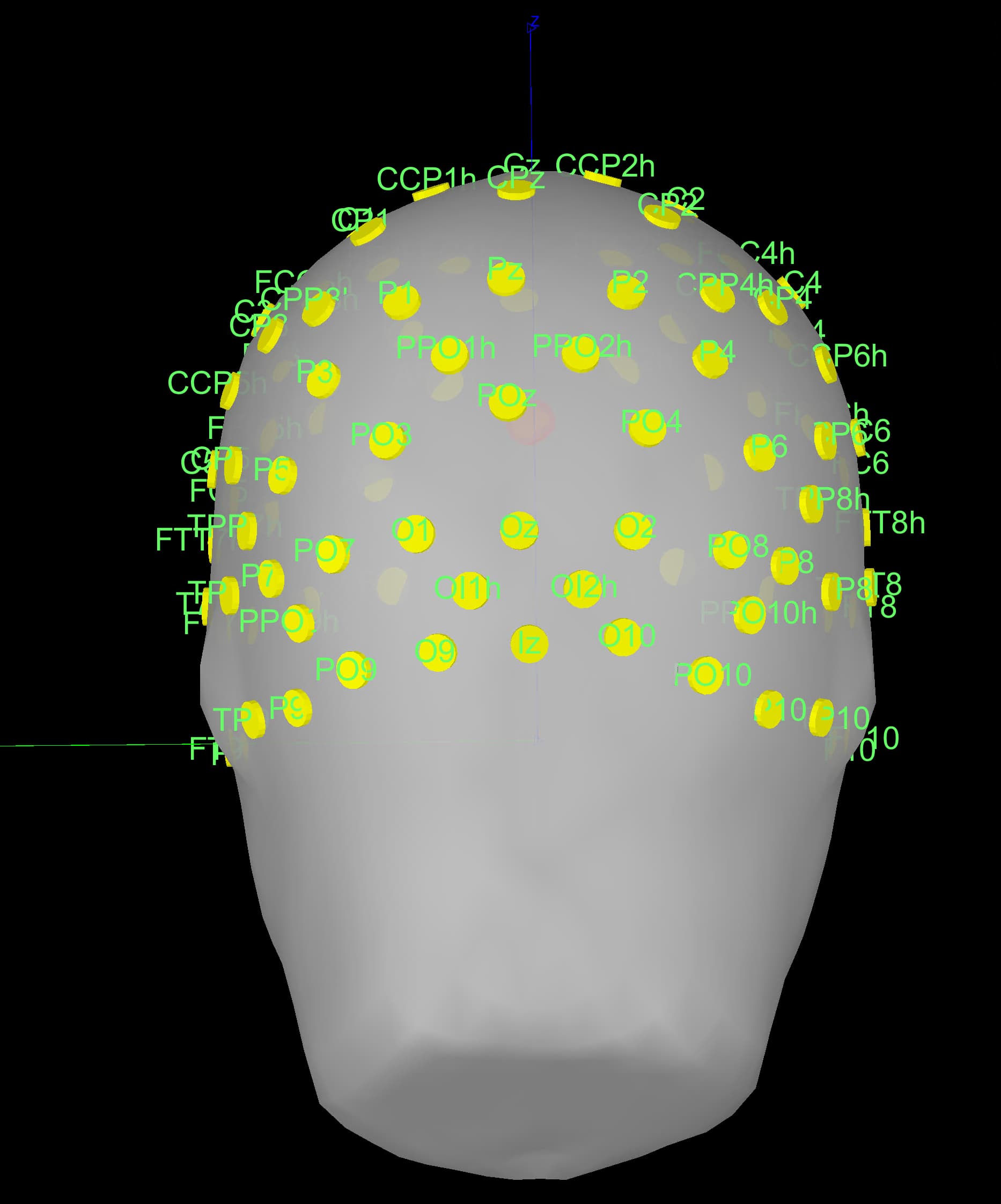
How do I know that the fiducial points are well-aligned in the 'MRI check' figures above? The LPA/RPA (y) axis seems to be slightly lower (maybe 1-2cm?) than what Brainstorm follows. Note: This image was not been edited at all after importing CapTrak positions.
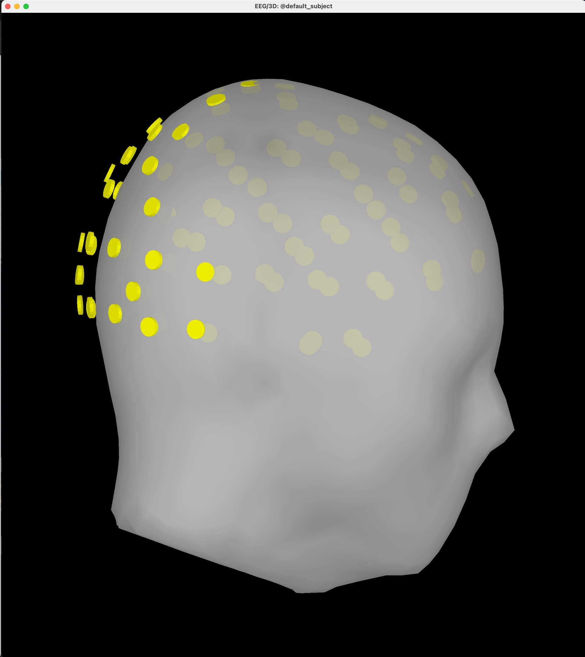
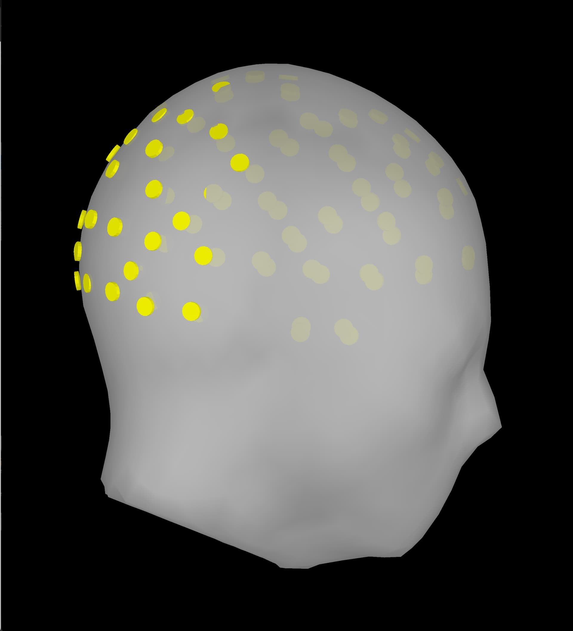
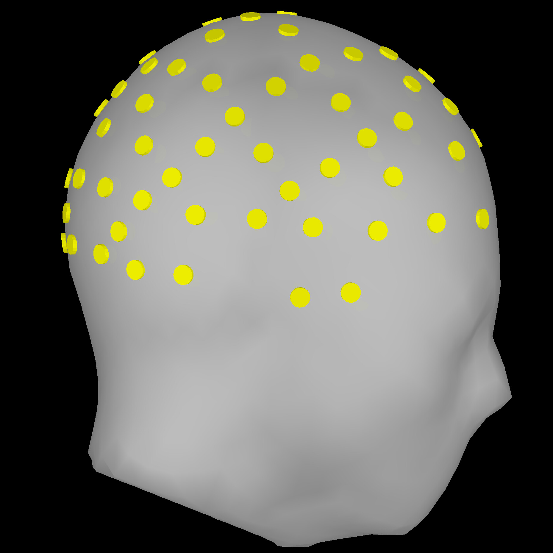
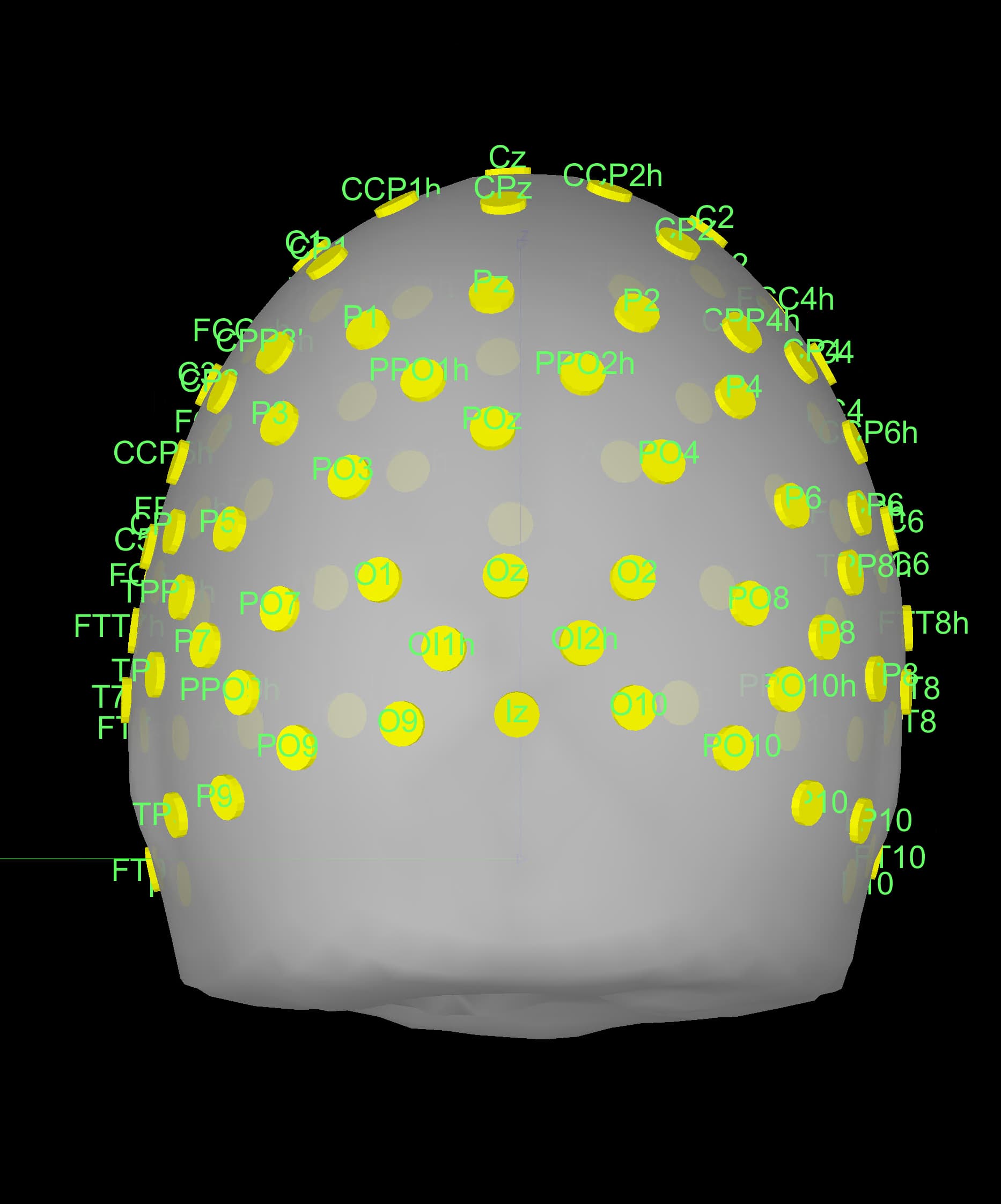
After projecting electrodes to surface to the images in my last post, the electrodes look like this on the default head:
Does warping anatomy potentially change the position of electrodes slightly? I'm trying to determine why I am seeing a difference in electrode positions between the CapTrak software's electrode position viewer and in Brainstorm. Specifically, there seems to be a deviation between the CapTrak software and Brainstorm in the posterior electrodes along the Z axis.
An alternative possibility is that the default Brainstorm fiducials and the data collection are slightly off between each other. Just trying to figure out what's going on. Thanks in advance!
Below are screenshots of (1) electrode positions before warping anatomy, (2) electrodes after warping, (3) captrak software electrode viewer. The only thing i did to screenshot 1 is project electrodes to the surface.
Please let me know if you need any additional information from me. Again, I appreciate the help!
It does not. Warping only creates a new set of anatomy files. These files are the Default anatomy files warped to fit the best the head points and sensors.
Did you warp the anatomy after projecting the electrodes on the head surface?
The head surface for figure 2 does not seems to correspond to the head surface for the Default anatomy showed in figure 1.
The deviation towards the right on the positions for electrodes Oz, Iz seems to be there in figures from both Brainstorm and CapTrak