Hello,
We have noticed a rather worrying discrepancy between MNI coordinates reported in BrainStorm on the one hand, and both SPM and Mango on the other hand. We used Colin27 preprocessed with BrainVisa (default template) in BrainStorm (version 3.1), as well as Colin27 from MNI website in SPM (version 5) and Mango (version 3).
In the first image, you can see on the left the BST MRIviewer, with the cross-hair targeting a well defined anatomical structure, with a report of MNI coordinates:
BST coordinates (MNI) -18.1 -35.1 -3.1
on the right, the same anatomical structure is targeted with Mango, the coordinates appear in the small window above BST viewer:
Mango coordinates (MNI) -17 -30 -7
ie a 5 mm difference along the y axis and a 4 mm difference along the z axis
In a second step, we loaded Colin27 in SPM5, targeted the same region and found the same coordinates as in Mango (image 2)
SPM coordinates (MNI) -17 -30 -7
Last, we entered the BrainStorm coordinates in SPM, to visualize the shift between the BST and SPM-Mango coordinates (image 3).
Any comments on this issue would be appreciated. Thank you very much!
Hyeong
Hi Hyeong,
Yes, it looks that something went wrong with one of the updates, at some point.
I’ll address this problem soon, I’ll keep you posted.
Thanks for reporting the issue.
Francois
Dear Hyeong,
I finally have time to review in details this problem that you exposed, sorry for the long delay.
After checking in details the procedure I’m using to calculate those MNI coordinates on the Colin27 brain, I found indeed some problems relative to the very definition of what the “MNI coordinates” are. I’m going to fix them with the help of Prof Louis Collins later this week. I also understood that there are some concerning issues with the way SPM/Mango generate those “MNI coordinates”, we are probably both wrong.
In the meantime, I think there are already a few confusing points we can clarify:
-
The template brain in Brainstorm is currently based on FreeSurfer, not BrainVISA. Please update your Brainstorm version.
Then create a new protocol to see the new defaults, or use the popup menus Use template > Colin27.
-
[removed because incorrect]
-
To help us understand where these differences are coming from, could you please do the following:
Thanks,
Francois
Hello,
We are replying on point-3.
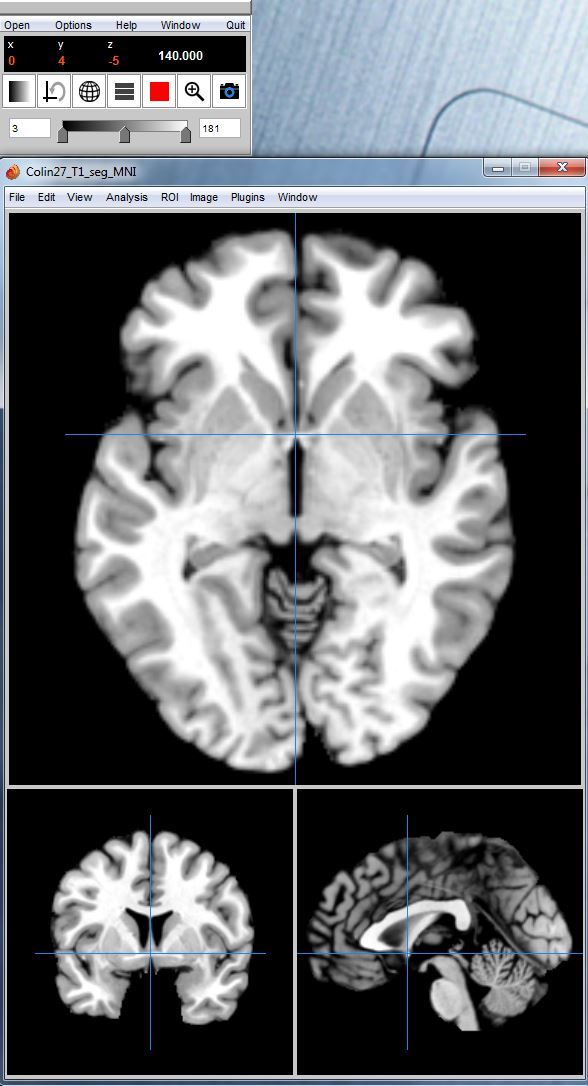
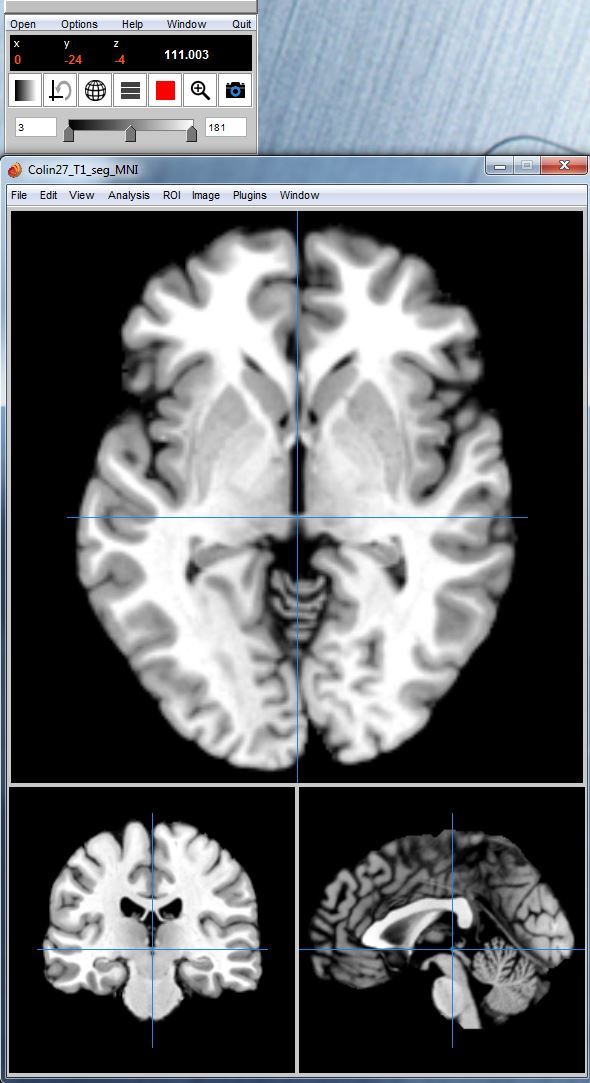
We report the MNI coordinates in Mango: AC(0,4,-5), PC(0,-24,-4). From what we read, it was expected that AC is not (0,0,0) in MNI space, although AC is (0,0,0) in Talairach space. We post the corresponding screen shots: mango_AC.jpg, mango_PC.jpg.
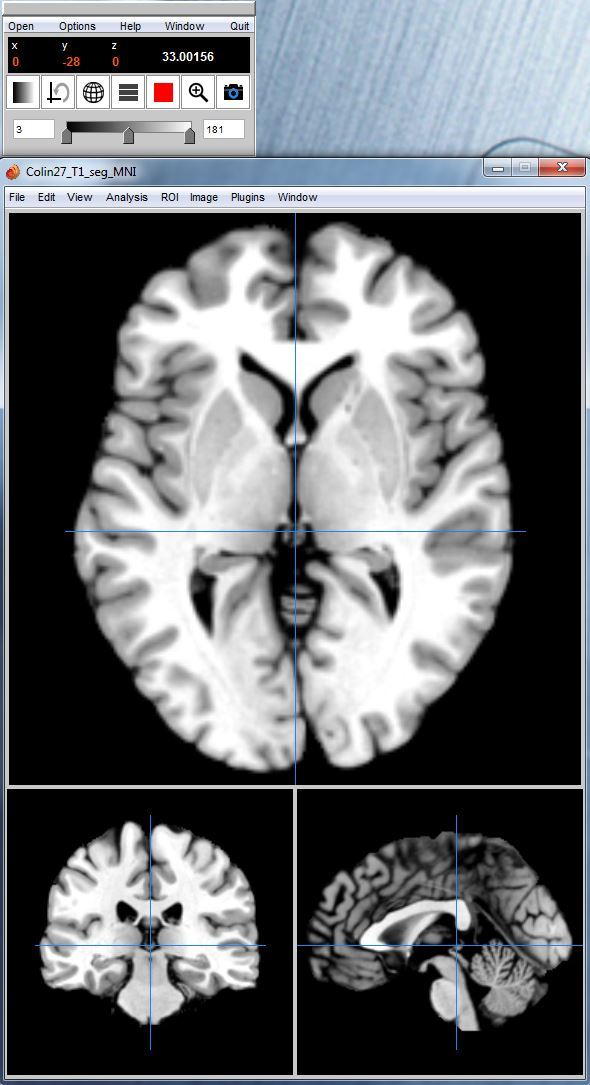
We also post screen shots for MNI (0,0,0) and (0,-28,0) in Mango (exactly same pictures in Mango and SPM): mango_0_0_0.jpg, mango_0_minus28_0.jpg.
Thank you for following this up.
Hyeong
Dear Hyeong,
I updated the way those “MNI coordinates” are managed in Brainstorm:
http://neuroimage.usc.edu/brainstorm/CoordinateSystems
For the MNI template brains (Colin27 and ICBM152), I tried to make them as close as possible to what we get when opening those volumes with the “Display” program from the MINC tools:
http://www.bic.mni.mcgill.ca/ServicesAtlases/Colin27
http://www.bic.mni.mcgill.ca/ServicesAtlases/ICBM152NLin2009
http://en.wikibooks.org/wiki/MINC/VisualTools/Display
It should be closer to what you’re observing in Mango/SPM, but might not be exactly the same.
Ultimately, those coordinates are valid as a normalized coordinate system only when working with volumes that have been transformed to the MNI stereotaxic space (this includes the SPM normalization). On individual brains they are just one subject coordinate system, but cannot be used as a normalized reference.
As I don’t want to risk any hazardous conversions anymore, I disabled the “MNI coordinates” display for all the brains that are not either the Colin27 or the ICBM152 templates.
The proper way to localize the position of a brain activity pattern in MNI coordinates is to 1) project the source results on the template brain and 2) reading the MNI coordinates from this projected dataset.
To use this properly, you have to update Brainstorm and update the MNI template you are using in your protocols, as described in my previous post.
Thank you for your reports. Please let me know if you have further questions.
Cheers,
Francois
FYI: There is now the possibility to compute MNI coordinates for individual subjects:
http://neuroimage.usc.edu/brainstorm/News#MNI_coordinates
The volumes exported from Brainstorm in .nii (T1 and sources) are now aligned in a different way, centered on the MNI coordinates (0,0,0).
The way they are displayed in SPM/Mango/MRIcro may change significantly.