Hi, First of all, I would like to thank you for your guidance in topic "head modeling" , then
i wanted to ask you that if i want to do EEG inverse problem myself (brain source signal estimation), can i display the estimated source signal with Brainstorm, i know that if Brainstorm does source estimation itself, it has the item "display on the cortex" for displaying the results, but my question is that: is it possible to import current signals and then use Brainstorm just for displaying them on the cortex ?
thanks
if i want to do EEG inverse problem myself (brain source signal estimation), can i display the estimated source signal with Brainstorm
Two options:
- Run the computation from a Brainstorm process, see this tutorial, in particular the beamformer example: https://neuroimage.usc.edu/brainstorm/Tutorials/TutUserProcess#Examples-1
- Compute a template source file in Brainstorm ("Full sources"), then edit the .mat file and replace the ImageGridAmp variable. Most of the information you need are in the Scripting tutorial:
https://neuroimage.usc.edu/brainstorm/Tutorials/Scripting
See the source estimation tutorial for a full description of the source file structureshttps://neuroimage.usc.edu/brainstorm/Tutorials/SourceEstimation#On_the_hard_drive:
Hi
i want to ask you a question about displaying on cortex.
as you said before for displaying source signals on cortex , i edit the parameter "imageGridamp".
but when i want to estimate sources in MUSIC method, i have just the correlation as you know, so how can i show the source location on cortex or MRI view?...
i estimate the correlation of all voxels and the vector with dimention(Nvoxels * 1) was created, the voxel with maximum correlation has a big chance of being source, when i edit the imageGridamp and changed it with this vector and click on "cortex view" i encounter with error.
thank you for your guidance.
how can i show the estimated source voxel with Brainstorm?
If you modify the number of time samples in ImageGridAmp field (the second dimension of the matrix), you must edit the Time field as well.
See this discussion:
thanks for your answer...
now, i have another question about displaying signals on the cortex from you.
i create head model with brainstorm with 20002 voxelnumber, and then i solve inverse problem with my method and then i wanted to show the estimated voxelsignal with brainstorm, so i have a matrix with dimention (20002*500) for voxels signal, after editing the item "imageGridamp" ;and click the item MRIviewer, the below picture has been shown for me:

as you see,the voxel with the big signal shown like dot!!!!!!!!! very little dot...It is very difficult to find them and distinguish their colors and the difference between their colors.... i wantet to have picture like below:
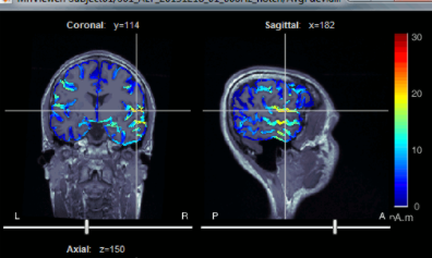
so what is your suggestion for me to have a good picture for article?
thans
Make sure there is no threshold applied in the Surface tab of the main Brainstorm window.
i dont understand your answer, can you explain more about it?
how can i modify the image grid map for my source signals? now, i have another question about displaying signals on the cortex from you.
i create head model with brainstorm with 20002 voxelnumber, and then i solve inverse problem with my method and then i wanted to show the estimated voxelsignal with brainstorm, so i have a matrix with dimention (20002*500) for voxels signal, after editing the item "imageGridamp" ;and click the item MRIviewer, the below picture has been shown for me:
as you see,the voxel with the big signal shown like dot!!!!!!!!! very little dot...It is very difficult to find them and distinguish their colors and the difference between their colors.... i wantet to have picture like below:
so what is your suggestion for me to have a good picture for article?
thans
Please consult the source visualization section of the following tutorial chapter:
https://neuroimage.usc.edu/brainstorm/Tutorials/TutSourceEstimation
@haniehsotudeh
What did the file you edited look like when displaying it in Brainstorm?
I guess this is not what you posted as your second screen capture...
Please post screen captures of the file displayed in Brainstorm before and after your modification.
If these are supposed to be surface-based results, please post images of the surfaces, not the MRI viewer.
Hi Frocois
i have ask you different question
if i wanted to see the result of MUSIC algorithm in MRIviewer,before you guide me about average the file and then edit the imagegrid amp,but if the averagind inactive for the file, how can i show my result MUSIC method in MRI viewer?
I'm not sure I understand your question.
Please create a new topic on the forum for each new question, and include some more context describing what you are trying to do.
1 Like
Hi~I want to visualize the statistical analysis results on the cortex and apply color mapping, but my output only includes the p-matrix and t-matrix. How can I map the comparison results to brain regions for visualization? Cause I want to see the " Display on cortex"
