I've run into an issue digitizing EEG electrode positions. I can accurately digitize the fiducial positions, however, the digitization of electrode sites seems to get warped somehow. I have successfully digitized several participants (E.G., image EEG_3D_02) using my current setup, but now I consistently get strange looking results.
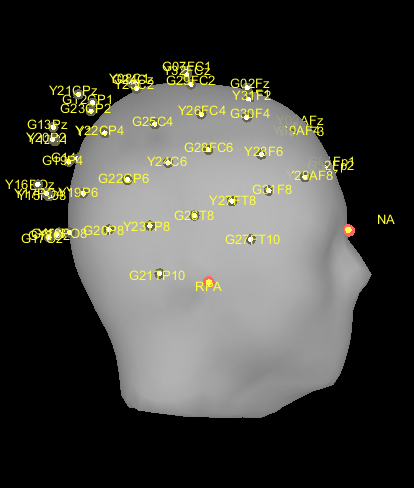
Here's a head I digitized last week.
(There's meant to be an image of a successful, normal looking digitization here, but the forum won't let me have two images...)
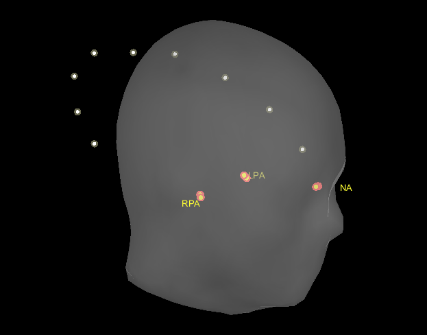
And here's a series of medial electrode sites I digitized using a dummy head as part of my troubleshooting yesterday.
It should approximate a medial view of the head, but you can see the shape produced is way off.
I take extra care to ensure the receiver (mounted on a set of glasses) stays still relative to the head position so I'm not sure what might be happening.
I'm using Brainstorm version 12-March-2024 with MATLAB R2023b and the Fastrak system.
Any advice would be tremendously appreciated - happy to provide more information as needed.
Have you tried to plot the digitized positions (.pos file), before importing it in Brainstorm?
To verify their position
Could it be that the fiducials in the anatomy were edited?
Right-click on the MRI for the subject, then Edit MRI... and View the positions of NAS, LPA and RPA.
I noticed that the nasion fiducial in your figure is just called NA, was it called like that when it was working well?
Yes, I am using the Brainstorm Digitize application.
I'm afraid I'm not sure what you mean in your second question. I use the Brainstorm Digitize application to record the spatial coordinates of electrodes and I can tell the coordinates of the electrodes are off by looking at the viewer as I record each electrode site. The images I upload are screenshots of the digitizations loaded into the viewer from our Brainstorm protocol.
Unfortunately, we don't have MRI for our subjects, so we are just using the default anatomy. We have not edited any of the fiducials.
I think the nasion has always been labeled NA in the viewer for us.
Here is the image I mentioned previously of a successfully digitized subject using the same setup.