Hi all, I have searched the forum as well as a plethora of site in order to get an answer and am at a loss.
My question concerns the removal of a stimulation (Grass S88 stimulator) artifact appearing as the result of a median nerve stimulation in order to evoke SEP of the contralateral somatosensory cortex. The artifact spans over a broad frequency spectrum and as such filtering is more or less out of the question.
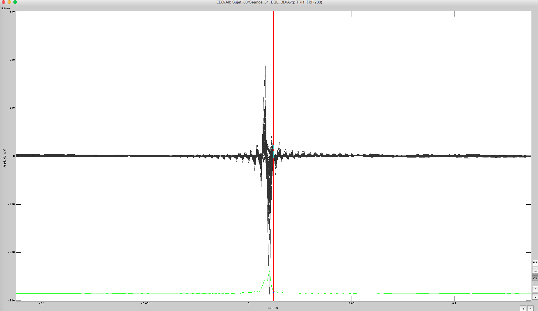
I have attached the worst case scenario in an image below and would be grateful if any of you had an idea as to what are my options. Again not all participants look like that and for some of them, the process: "cut stimulation artifact" succeeds in removing the annoying artifact. Then again for some of the participant this process is not sufficient in order to do so
Thanks again in advance.
Hello,
There are filters already applied on this file. It is impossible otherwise to observe the stimulation artifact before the time of stimulation.
So the first thing to do: do not apply any frequency filter to your recordings (you may have to change your export or acquisition parameters).
Then, if you are not planning to do any time-frequency decomposition of your trials, you could remove the stim artifact with the process “cut stimulation artifact”.
This process replaces the recordings with a linear interpolation at the times you indicate in the options. This introduces severe artifacts in the frequency domain, so it is mostly for visualization purposes that you would run this process.
It does not “remove” the artifact, it just masks it so that the default scaling in the figures look good.
The last thing you could try is to extract ICA components for the artifact.
Feed your entire file to the process “ICA decomposition” and try to find one or several components that remove your artifact.
(sorry, this process is not documented well yet, please seek for help in the EEGLAB documentation)
http://neuroimage.usc.edu/brainstorm/News#March_2015
Cheers,
Francois
Hi François
That is also what I was thinking (filter already applied). However I tried to locate where and how these filters would have come into play and I can’t locate them anywhere. I am using an EGI netamp 300 and my acquisition setup does not contain a filter. Moreover, export of the file have been performed without the application of any filter. Even tough I am quite aware of the fact that your specialty is the end-user software, I was wondering if you have ever encountered something similar.
I am tempted to call EGI directly in order to find out if there is an analog filter somewhere that could cause this behavior.
thanks in advance
Jean-Daniel Dubois
Hi Jean-Daniel,
Indeed, you can contact the EGI customer support and ask them to explain why you observe this strange behavior.
However, the screen capture you sent us may not be very meaningful to them, as they would primarily suspect some post-processing done in Brainstorm.
Can you reproduce the same type of the display directly in the NetStation ?
Francois
Well yes and no.
I can definitely see the artifact in Net Station but the screen capture is from the averaging of multiple epoch in Brainstorm.
I already spoke to their customer support and I am wondering if an anti-aliasing filter in the amplifier (confirmed by their tech) could be distorting the artifact as much.
Thanks again
If this the most unprocessed data you can get, I’m not sure how you could recover any brain signal of interest underneath…
You have to understand why your EEG system recorded this, so it doesn’t happen again in the future.