Hello,
I am newly learning brainstorm.
Thanks a lot for your clearly tutorials.It really helped me a lot!
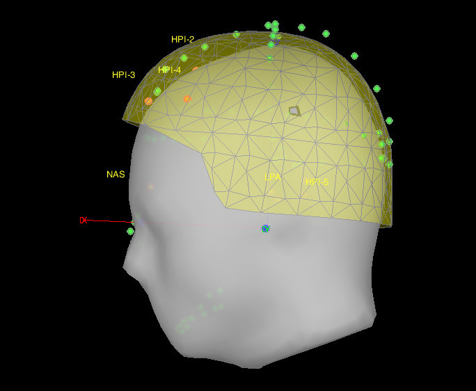
Since I don't have the individual anatomy file, I used the templates. However, most of the participants's MRI registration view is like the picture shows. The green points are just on the outside of the cap. Have you encountered the situation like this and do you know how to fix it? Thanks in advance!
If you observe some of the green points (head shape digitized with the Polhemus) outside of the MEG helmet, there is something wrong with the registration between the Polhemus points and the MEG. This is independent from the anatomy you use (template or individual MRI). You are maybe not pointing a the correct anatomical locations when doing the digitization of the fiducials and head shape.
Which MEG system is this? What software do you use to digitize the points?
After you solve this issue, you will may need to align better the MEG sensors with the MRI. Your nasion marker looks very high, in the middle of the forehead…
If you are using a template anatomy, do not try to use the automatic registration, because the shape of the head does not match the shape of the digitized points.
Sorry for my so late reply. The system we used is Elekta Neuromag, and we used Isotrak 3D digitizer (Polhemus) and acquisition do the digitization. During the data analysis, ICBM152 anatomy template has been used, I don't know if this is the main reason for the problem, or because we also digitized some point below nasion.
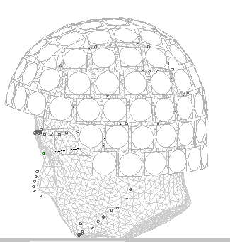
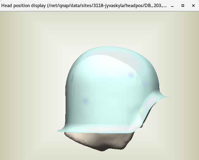
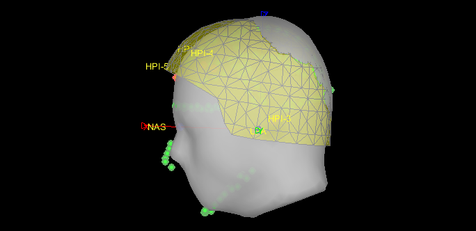
But now there is some more problems pumped out. Now after we import data into the brainstorm and the MRI registration will be shown like the first picture below, the helmet seems somehow cutted into the brain and there is no similar problem happened in Xfit and DACQ workstation (like the second and third picture below). We don't know if this is caused by some display bug or other issues. Have you met this kind of report before and do you have some suggestions to fix it?
First, the helmet displayed in Brainstorm (the yellow transparent surface) is an approximation of the helmet surface, not an accurate model like the one you should in the bottom picture, so it is normal if you don't see the same thing in the two programs.
It also looks like your digitized head points are lower in Brainstorm: The nose and chin points are clearly much lower in your Brainstorm figure, so the registration you use is not the same. This could be due to two things: the position of your NAS/LPA/RPA points in your MRI in Brainstorm is not correct (as compared to the ones you defined during the MEG acquisition), or you modified this registration in Brainstorm after importing the recordings (never use the option to "Refine registration automatically" when using an anatomy template or with less than 100 head points).
Hi, Francois
Firstly, thanks a lot for your kind help and all those detailed tutorials on the website. It really helped me a lot.
For the second suggestion, the picture I have here is the original figure I got when I imported my data into Brainstorm. So I didn't modify it yet.
And for the first one, I thought it might be the key point but I didn't find a really good solution to fix it yet. As it mentioned in the tutorials, it's really hard to indicate those points with a MRI view. So do you have any suggestions also for the coordinates of these anatomical points in MRI vies as the example gave below? https://neuroimage.usc.edu/brainstorm/CoordinateSystems#MRI_coordinates
And for the first one, I thought it might be the key point but I didn't find a really good solution to fix it yet. As it mentioned in the tutorials, it's really hard to indicate those points with a MRI view. So do you have any suggestions also for the coordinates of these anatomical points in MRI vies as the example gave below? https://neuroimage.usc.edu/brainstorm/CoordinateSystems#MRI_coordinates
You are supposed to place the fiducial points NAS/LPA/RPA in the MRI using the same convention that you used for the MEG acquisition. Only you can decide where this points are supposed to be.
I think it is useless to spend too much time trying to find the perferct points: if you are using an anatomy template, this registration is necessarily wrong anyway...
If you had access to more digitized head points, you could warp the template anatomy to match the shape and size of your patient: https://neuroimage.usc.edu/brainstorm/Tutorials/TutWarping
To keep in mind for your following acquisitions...
Hi, Francois,
Thanks for your kind suggestions, and after I warped the template for my participants, the results looks much better now.
Now I'm going to do the group analysis, and I noticed that in turorial you suggested to do the subject coregistration to reproject the sources of all the subjects on the default anatomy. However, for my study, I don't have individual MRI data, but warp the templet MRI based on the digitized points, do I still need to coregistartion them?
don't have individual MRI data, but warp the templet MRI based on the digitized points, do I still need to coregistartion them?
No, if you have source maps estimated on the warped cortex surfaces, you should be able to average and compute other statistics directly across subjects without any additional step of projection.
For organizing better your database, or if some processes complaining about multiple subjects, you could also decide to project first your individual source maps on the default anatomy. This would simply rewrite the source maps pointing at the cortex surface of the template, without doing any additional interpolation.
The two approaches should give you exactly the same results.