Project electrodes with MNI fiducials visible

Hi Brainstormers,
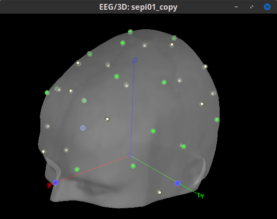
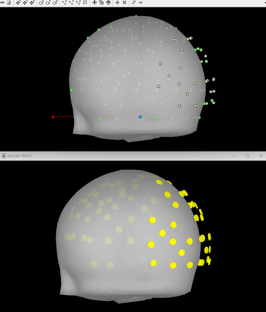
I am wondering if it is possible to project electrodes to the 3D head model generated directly from the standard MNI brain, after clicking this button (green circle):


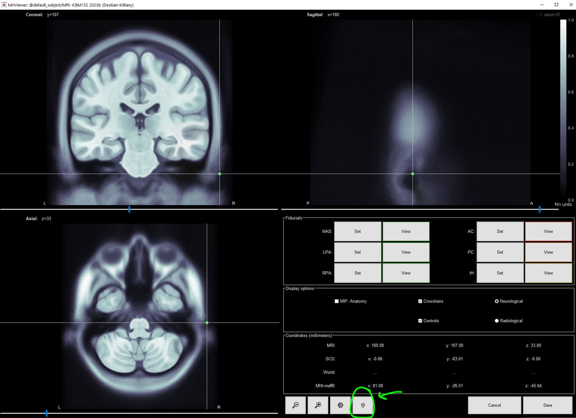
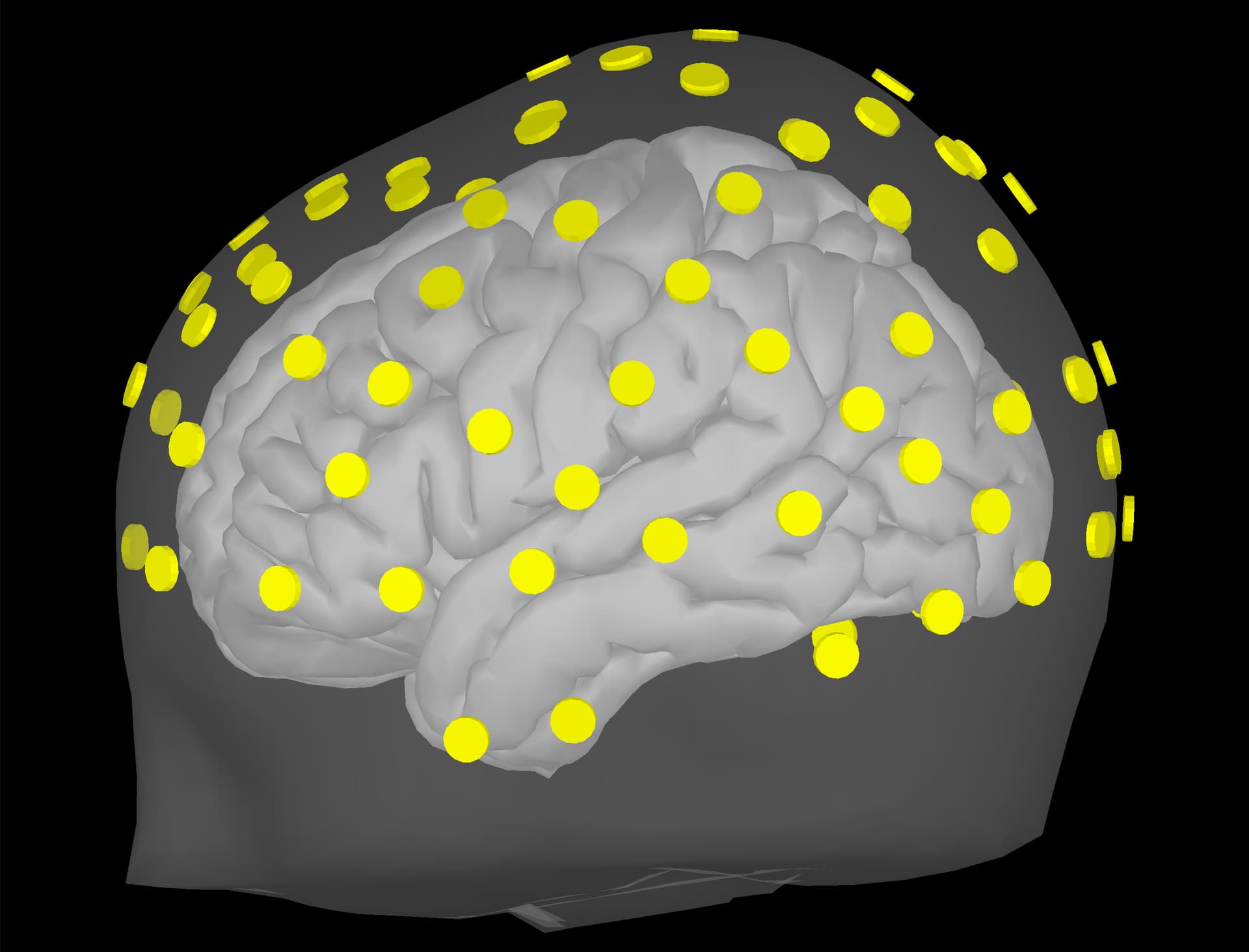
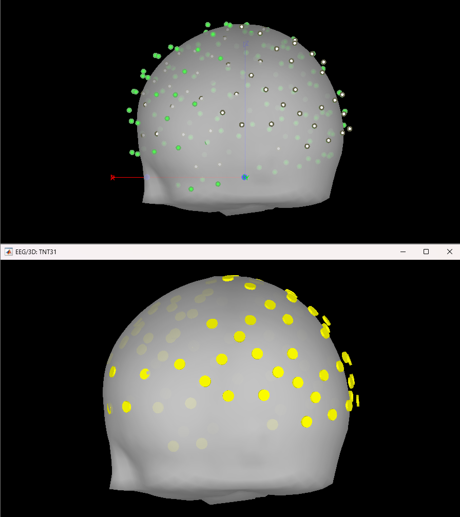
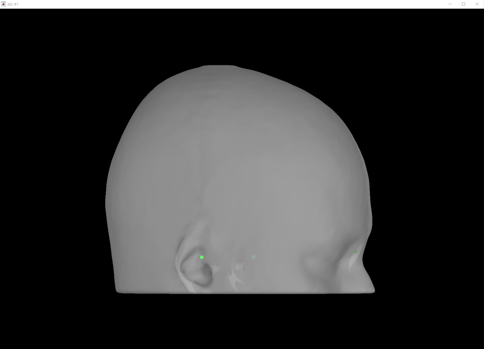
I ask because when projecting to the BEM head model, the fiducials of the MNI brain (which Brainstorm provides) is not very clear to me. I like the visibility of these green dots in this 3D reconstruction:

Maybe there is another way to do this?

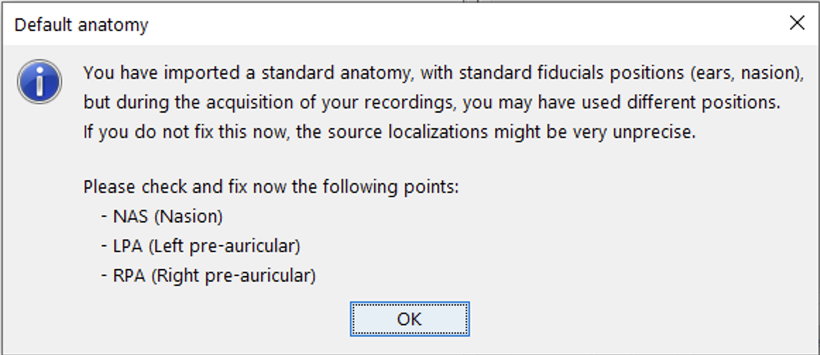
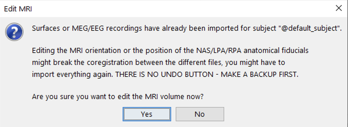
I am inquiring about doing this this because I'd like to ensure that there is an appropriate registration of fiducials between the standard MNI and our CapTrak fiducials, or if I need to edit the standard MNI fiducials. I feel that they are going to be pretty similar, but I want to ensure of this. If there are any differences, I would say that perhaps the RPA position is a little far backwards (caudal) compared to our positions (visible in screenshot 1). I thought we had identical positions until I double checked after seeing these prompts:


How much measurement error (in mm?) between the 2 sets of fiducial points is "acceptable"?
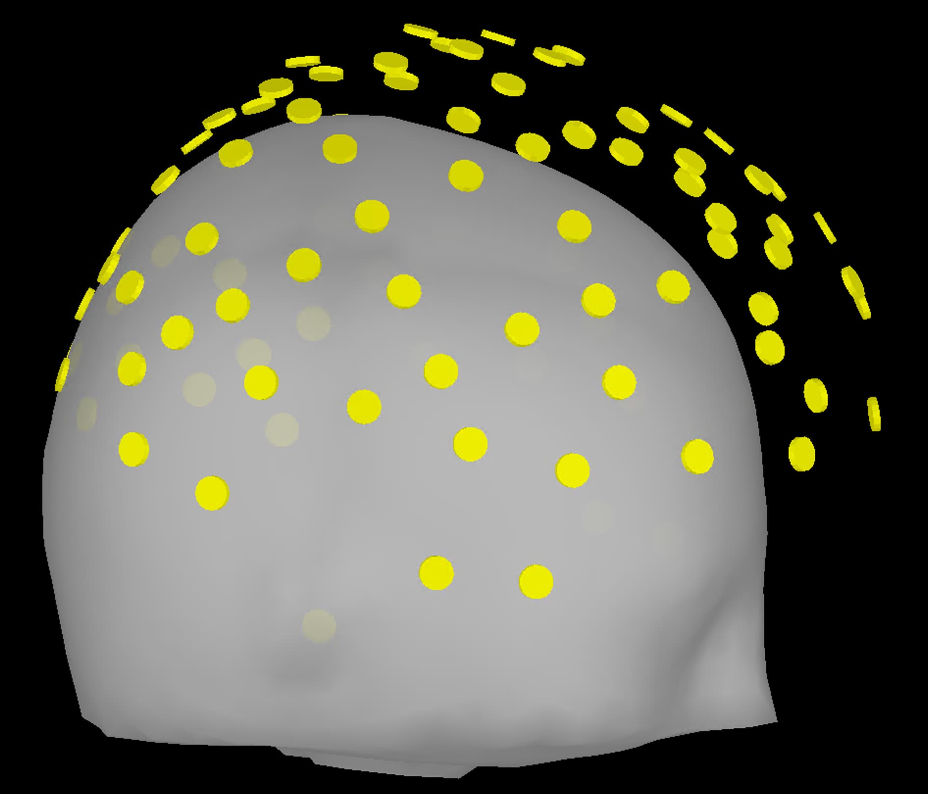
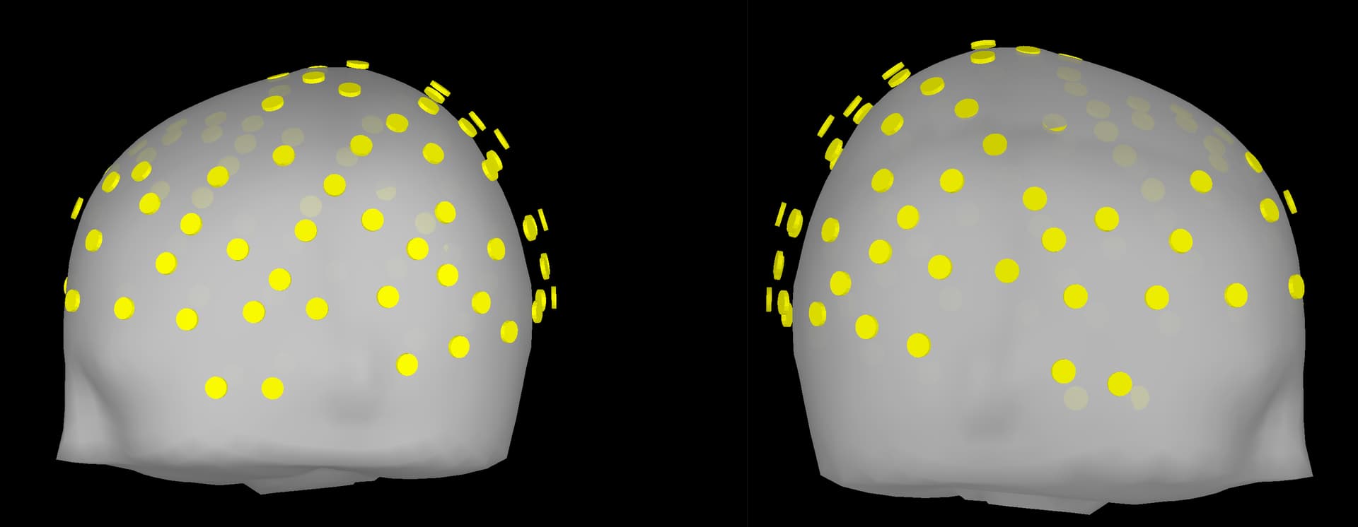
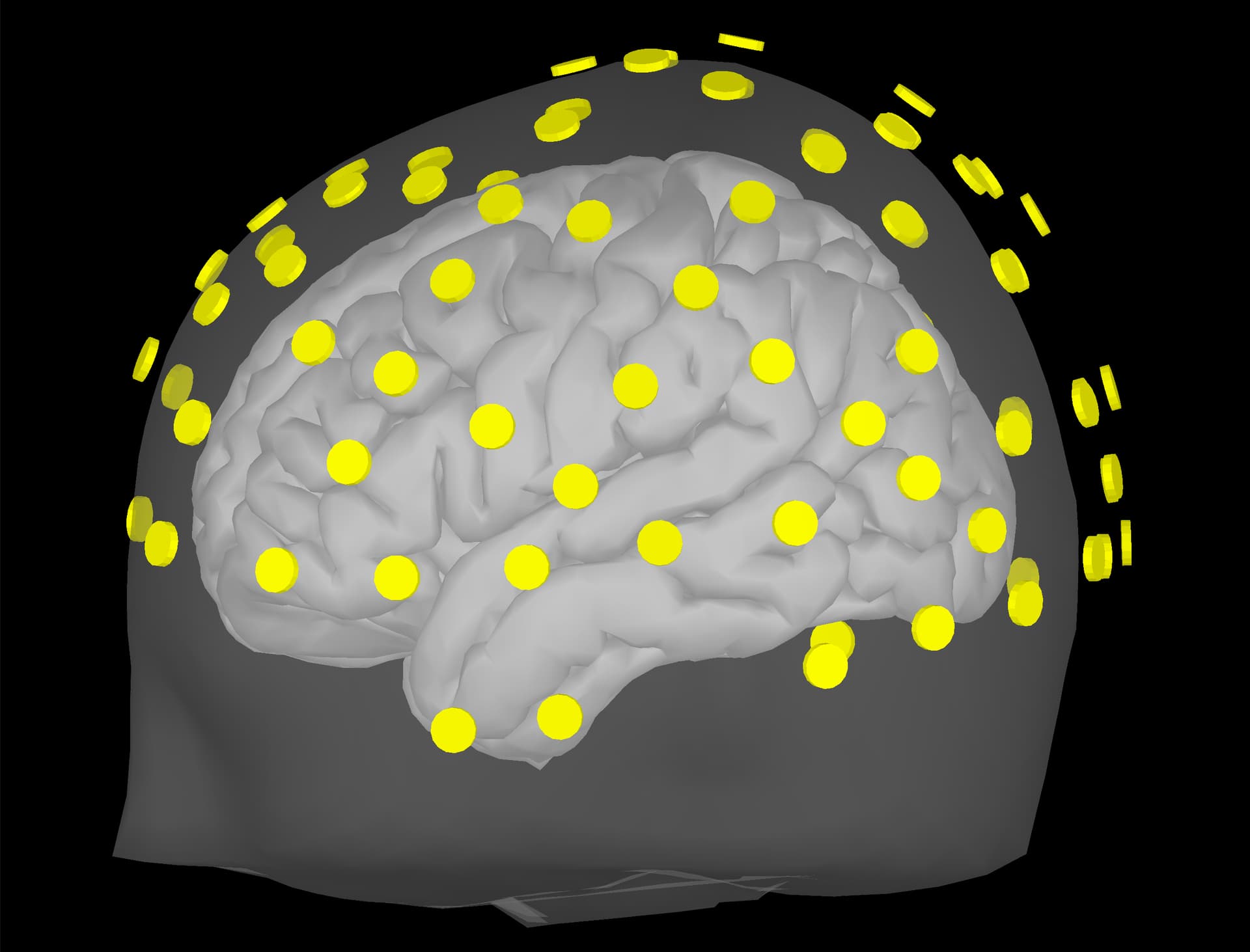
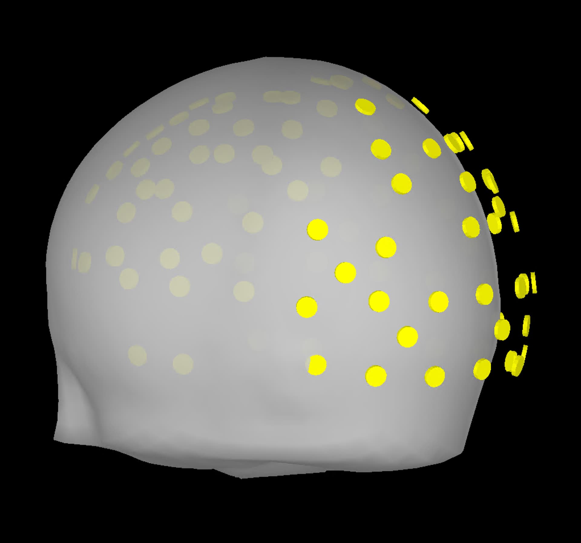
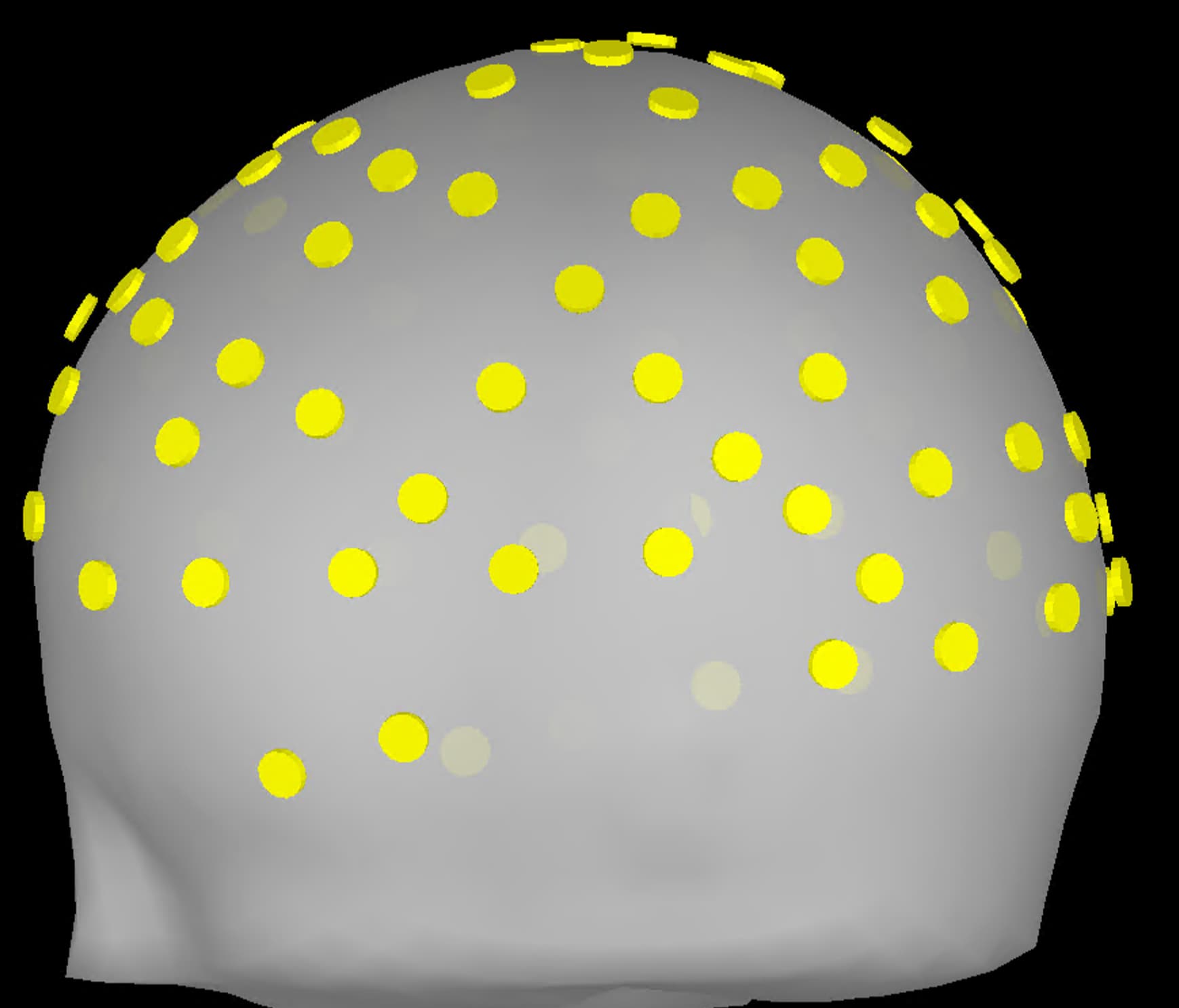
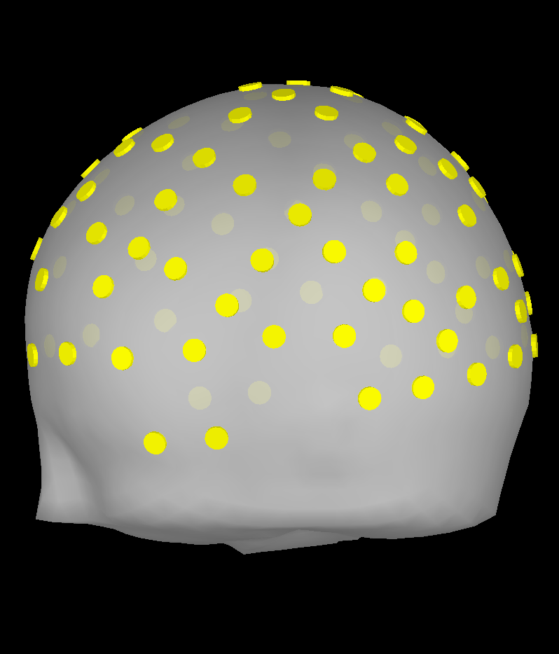
Upon importing CapTrak positions onto the standard MNI brain, I obtained this result (after projecting electrodes to surface), which look completely reasonable to me.

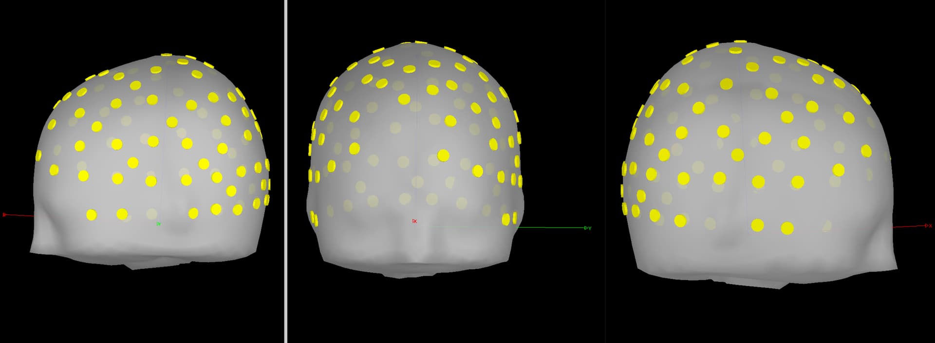
However, I'm wondering if this visual inspection is robust enough to ensure quality EEG-MRI registration with the MNI brain. In looking into File Contents of the CapTrak data, I also notice that the fiducials for CapTrak and MNI brain positions seem slightly off, but this isn't seemingly present in screenshot 5. For example, File Contents of CapTrak give these coordinates (which were in m that i converted to mm):
NAS: [113.2, 0, ~0]
LPA: [13.5, 88.8, 0]
RPA: [13.5, -88.8, ~0]
whereas the MRI viewer of MNI brain told me the following (in mm):
NAS: [101.6, 0, 0]
LPA: [0.66, 83.01, 0]
RPA: [-0.66, -83.01, 0]

While of course there are differences, I am wondering if I am needing to change the positions of the MNI fiducials.

Thank for you for your time. I greatly appreciate your expertise and advice.

Sincerely,
Adam

Just wanted to add on something about this:

The above screenshots projected to the electrodes are in contrast to something like below, where the electrodes, for some reason, seem to be off position. I wonder if these apparently misplaced electrodes are presenting like this because of incorrect LPA/RPA/NAS landmarks, or because perhaps the head shape is different between this subject from which we obtained the CapTrak data and the MNI head.

In the latter instance (different head shapes) would it be permissible to project the electrodes to the head and proceed with processing?

Thanks again,
Adam

You can right-click the channel file, then select MRI registration > Check

  • Blue dots are the fiducials from the MRI
  • Red dots (if any, are the fiducials from digitizer)
  • Green dots are head points
  • White points are sensors

@Marc.Lalancette, any thoughts on this question?

These differences may rely on the fact that they are from two different coordinate systems. When acquired fiducials are located in the channel file that is imported in Brainstorm. The best fit between the two sets of fiducials (MRI and digitized in channel file) is computed and the transformation is applied to the sensors as well.

Have you tried to warp the Default anatomy to match the subjects head shape (from the digitized head points)? Fitting the Default anatomy to the head points seems a better plan than fitting the head points to the Default anatomy

https://neuroimage.usc.edu/brainstorm/Tutorials/TutWarping

Hello,

I would first echo Raymundo's comment that you should first warp the default anatomy to the rough size and shape of the participant's head, ideally from a set of digitized head points, but if you don't have those, you could try with the electrode positions themselves and see if it gives a reasonable shape (it doesn't always). After that, if you eventually do group analysis and warp to the template again, you would transform your source space results later, but those would first be computed with the best approximation of the participant anatomy.

Otherwise, you must realize that the correspondance between the default anatomy and your real participant anatomy will be poor, regardless of how "well" you align fiducials. And of course, if you placed the MRI fids on the default anatomy (not warped to head points), it's a different head size/shape and it won't match your real digitized fids. In that context, there is no recommended maximum distance between fiducial sets.

Furthermore, the MRI fiducials are used to define the subject coordinate system, and also to provide a first alignment with the functional (EEG/MEG) fiducials. But you can further align using the head points or manually. The important result is that the EEG/MEG fiducials (and electrodes) are in the right place on the head surface. The final position of the MRI fiducials is mostly unimportant and both sets don't need to match.

Hey Marc & Raymundo,
Thank you both for your thoughts. I appreciate it!

We have CapTrak, so I’ll try warping the default head to match the CapTrak electrode locations. Should warping be done for every patient using the default head, or only when there’s a noticeable mismatch between the CapTrak electrodes and the default head shape?

So far, I’ve projected CapTrak electrodes to the head surface (without warping) when the head shapes appear similar based on visual inspection. However, in cases like my latest screenshot, I was second guessing how to proceed. It seems possible that fiducial placement during data collection was off, or as we discussed, the head shapes differ in a way that a simple electrode projection can’t (or shouldn’t) try to correct.

Are you saying that (1) the coordinates listed in the File contents of the CapTrak data are in different coordinates than SCS, or (2) that transforming raw CapTrak RAS coordinates to SCS introduces some error? In File contents, I pulled the coordinates under the "-SCS:" listing, so I thought it'd be the same as the default fiducials, which came from the SCS coordinates listed in the MRI viewer.

Okay, this is great. Now understand that Brainstorm computes the alignment of fids between the two systems (default & CapTrak) as best as possible at import, automatically.

I think the main question I have now is my first question of this post: is it permissible to project CapTrak electrodes to surface when the head shape is similar? Or do I need to warp regardless?
Below is an example of a situation where CapTrak positions are similar to the default head (without me doing anything else).

My apologies for this turning into a novel. I appreciate the discussion, and thank you both for all your hard work for Brainstorm and helping us users. We really appreciate it.

Adam

Hi Adam,

The question is rather what's the best approach here. If you're interested in source localization, you want to approximate the real subject anatomy as best as possible. There's no reason not to warp, unless the warp result is worse (e.g. very distorted), or if your electrode positions are not that great (more on that below). In your last figure, the real subject head appears longer than the template by over a cm I'd guess. On the other hand, if your electrode positions are on top of somewhat large electrodes, and thus further away from the scalp, you should account for that as well before warping, or you'll end up with an artificially inflated head. The points you use should be on the subject's scalp surface. I'm not aware if there's a way to account for electrode height in Brainstorm, but I wouldn't think so, right @Raymundo.Cassani? Usually we always try to get head points in addition to electrodes. Some groups even digitze the head separately before putting on the EEG cap.

Again though, even with a lot of effort, this will still be just a template and any source reconstruction should be interpreted accordingly. For example some source modeling options should be considered, e.g. unconstrained or "loose" source orientations.

To clarify how the initial MRI-EEG alignment is done: Brainstorm will assume that both sets of fiducials are meant to define the same coordinate system. In other words, it doesn't match fiducials themselves, but creates the SCS coordinate system (based on the fids - see the coordinate system info in the tutorials) on each side independently, and then assumes it's the same coordinate system. But I wouldn't spend too much time placing MRI fids on a template, and instead "edit" the coregistration (where Raymundo pointed to earlier), to make sure your EEG fids and electrodes appear where you expect them to on the head surface.

If you have the head points for the subject, why not to warp. In the case that the subject has a head that is almost the Default anatomy, it will just happen that warping is close to nothing. This will help your pipeline, so all subjects are processed the same.

Thank you both for your time.

@Marc.Lalancette, could you please clarify what you mean by this? I am not sure I understand if this applies to our data or not. Are you suggesting that I exclude >2% of head points from the warping process (the default % in the warping tutorial) if I notice excessive # of head points for from the head? If there are a lot of head points far from the head, how do I know what % to exclude from the warping?

In the meantime, I tried warping with this participant and the results are below. They look reasonable, and from the tutorials, an "alien" head after warping is acceptable. What do you think? I used the default 2% head point exclusion. These electrodes have not been projected to surface, I figured this view was more informative than projected electrodes.


Thanks again for your time.
Adam

I'm saying that when you measure the positions, for warping they should be on the scalp surface, not on top of an electrode that adds 1 cm distance from the scalp for example. Hope this clarifies.

Hi Marc & Raymundo,

I have another question that is related to this.
I warped the default head to one set of CapTrak coordinates (baseline EEG session) and got good results with baseline CapTrak.
However, when I look at the second session CapTrak data on the warped head (that was warped to Pre CapTrak data), I got the following results (screenshot below). Is this an issue, or should I just project the electrodes to surface and move forward? Note: this is a different participant than the screenshots from above.

Thank you, I appreciate your time.
Adam

How was the alignment before warping?

It is reasonable, it seems. Below are two screenshots. One before warping, one after.
Still only the default 2% of head points were excluded.

No warp:


After warp:

Just to rephrase:

  1. There are two sets of head points from different day (Session1 and Session2).
  2. Anatomy is warped with points from Session 1. This creates an individual anatomy
  3. The head points from Session 2, are not aligned with the anatomy

You may want to check the registration (right-click > MRI registration > Check) for sensors in Session1 and Session2. For Session1, the registration of the fiducial sets (anatomy and sensors) should be pretty good, as the anatomy was warped to match those points.

For Session2,

  • How is the registration between the anatomy points and the sensors fiducials?
  • Have you tried the menu (right-click > MRI registration > Refine using head points?)

Yep, correct.

It looks like the registration is decent for session 2, but refining with head points seems to fix the registration a bit - I think this may be the solution. Here are a few screenshots.

Just after import:


After refining electrodes with head points:

After projecting electrodes:

I think these are all the questions I have for now. Thanks again for the help Raymundo!

Adam

Hi Raymundo & Marc,

I have a follow up question pertaining to Marc's comment below and in the following tutorial: https://neuroimage.usc.edu/brainstorm/Tutorials/SourceEstimation#Unconstrained_orientations.

Our cohort includes people both with & without individual MRI data. Currently, I use constrained sources for those with MRI. Should I also use constrained sources for those with template MRI to maintain consistency, despite Marc’s comment and the tutorial stating it might be better to use loose/unconstrained sources when no MRI is available? Or is it preferable to follow their advice and use loose/unconstrained sources for subjects without individual MRI?

Thank you all!

When the default anatomy is used, a smooth template default cortical surface is used and it does not reflects the individual topography of the individual's cortex. Using constrained orientations, the dipole orientations are set to the normals of the cortex, but since this default cortex is not the "original" indidual anatomy, the orientation assumption too strict. Thus the use of unconstrained.
For the individuals, the cortical surface is derived form their MRI, thus this assumption is ok.

If you are planning to compare the results across subjects, it seems appropriated to use the same approach, thus differences, cannot be attributed to the different methods.

Raymundo,
Yes, we are doing group analysis treatment vs control. I was thinking I would need to keep constrained for all subjects so as to not introduce any differences between groups. Thus, I will have constrained sources for all subjects and acknowledge as a limitation.

Thank you again!

This introduces the issue of incorrect dipole orientations for subjects using default anatomy, as described in the tutorial, and Marc's and my comment above.

Raymundo,
I thought with what's below, you were suggesting that I keep the method consistent between all patients since I'm comparing two groups.

Since I use constrained sources for those with MRI, it seems reasonable to also use constrained source modeling for those without MRI—just to avoid introducing methodological differences between groups, even though constrained source modeling may not give very accurate dipole orientations without MRI.

Are you now suggesting something different from using the same (constrained) source computation method for those with vs without MRI? Or are you just adding context? I want to make sure I’m following you correctly. Thank you!

I was pointing towards using unconstrained orientations for all the subjects, with or without their individual anatomy.