Hi everyone,
We have been seeing a strange and persistent pattern in our EEG recordings and would like to hear if anyone has experienced something similar.
Setup:
-
Brain Products actiCAP + BrainAmp amplifier
-
Multiple cap sizes, matched to head size & sex
-
Participants: asian adults (Both Healthy and disease model)
-
Low impedances, controlled environment Previous system (Grass/Natus + Waveguard Cap, ANT)
-
resting EEG (eye-closed)
did not show this issue Problem Across all participants and experiments, we observe occipital-dominant activity in the topography, regardless of frequency bands.
It appears consistently even with different caps, operators, and recording days.
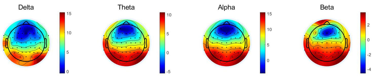
(an example of a patient, but healthy participants also showed same patterns)
While similar occipital activity can be seen in the high-frequency range with our previous Grass system, it is far less pronounced compared to the Brain Products recordings.
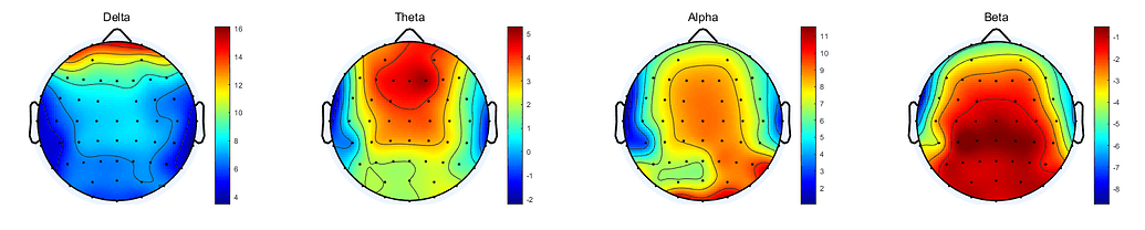
(Grass system, a patient case in the same cohort)
What we’ve checked Environmental artifacts → unlikely (appears in all subjects)
Alpha–beta misclassification → no change in alpha topography when redefined
Population-specific head shape → possible, but Waveguard data from same population was normal
Multiple preprocessing pipelines → same result
I’d like to know: Has anyone observed a similar occipital-dominant pattern with Brain Products systems? Any insights on possible causes (hardware, grounding, reference) or troubleshooting steps would be greatly appreciated.
Thanks,
Tae-Gon