Is it possible to import a CT with grid/depths and use that to create electrode locations with Brainstorm? I looked at a bunch of SEEG related posts and everyone seems to be importing the coordinates, if that is the case then what other software are you using to get the coordinates?
In Brainstorm, you can import multiple volumes (MRI or CT) for the same subject, but they must be registered with the MRI generated by the program you use to extract the cortex surface (same number of voxels, same dimensions, same orientation). We will improve this in the future, but for now it is sometimes complicated to work with multiple volumes (this software was not designed for this purpose initially).
You would be able to import a CT image on top of the MRI processed with FreeSurfer/BrainSuite, if you can get it in the exact same volume space. Then you would be able to mark your ECOG/depth contacts on the CT slices: http://neuroimage.usc.edu/brainstorm/News#February_2015
I am not very familiar with any clinical setup, so I don’t have any recommendation to make. Many users reported they were using the coordinates saved from their neuronavigation software, to which they were simply adding the same anatomical landmarks we use in Brainstorm (NAS/LPA/RPA).
Maybe you can try to contact other users who posted similar messages about SEEG/ECoG on this forum.
Actually, I realized that you can do more with Brainstorm than what I was mentioning earlier.
If the volume is registered in MNI space, and you computed an “MNI transformation” for the subject in Brainstorm, you should be able to visualize the two volumes simultaneously, and mark your contact positions using the CT.
First import the full MRI segmentation (“Import anatomy folder”) and compute the MNI transformation when setting the NAS/LPA/RPA points.
Then when you import the CT scan, select the option “Register”. http://neuroimage.usc.edu/brainstorm/News#MRI_coregistration
This solution involves the computation of two transformation with an anatomical template, therefore it would be less accurate than a direct registration of the subject’s MRI and CT. But it could help you, in case you don’t manage to register the two subject volumes otherwise.
hello there
we are working on a software called Gardel for finding semi-automatically the location of SEEG contacts, based on registration of CT with MRI, which will be integrated to our Anywave Software
[QUOTE=bowenseeg;11286]Is it possible to import a CT with grid/depths and use that to create electrode locations with Brainstorm? I looked at a bunch of SEEG related posts and everyone seems to be importing the coordinates, if that is the case then what other software are you using to get the coordinates?
In my hospital, skilled technicians mark and tag the locations in the CT, using in recent years CURRY, as part of the clinical process and for subsequent 3D visualization of the implantation. For bringing these CURRY data into Brainstorm, we wrote custom scripts to take the 3D coordinates from the clinical CT data (CURRY “POM” files), in the same coordinates as the scalp data from the CT (CURRY “skin” files). We then use the scalp points as virtual headpoints in Brainstorm, to register these headpoints to the scalp extracted from the MRI in the Brainstorm coordinates. The SEEG coordinates are then automatically in the same reference system as the MRI, without having to worry about precise coordinate definitions between the two software systems. We then finish the process by assigning the tagged locations to the corresponding channels and updating the Brainstorm Channel file.
We have been working to make this process a formal “Brainstorm” process (in your home folder), but are not ready to release it yet. The process may also be too specific to our combined Nihon Khoden / Neuromag / CURRY pipeline.
After loading the anatomy folder and computing the MNI coordinates, I imported the CT (convert to nifti first) same as an MRI.
I then chose to resample the CT.
After that I renamed my MRI#2 to CT and then I computed MNI coordinates for the CT.
Then, for the CT I did ‘register to default MRI’
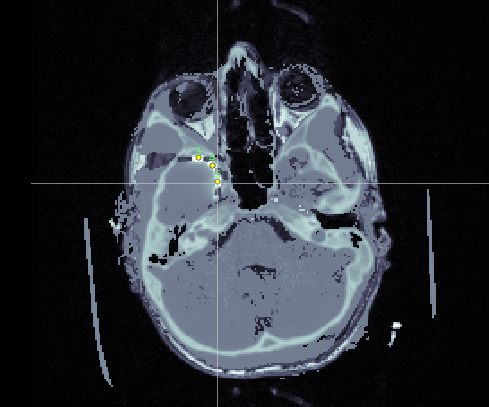
This process did fail a few times 'out of memory' error; it seems to use a ton of memory! I have 9GB on this workstation. I did walk away from the computer last night in a bit of frustration and when I returned this morning I had a CT_coreg there. To my enjoyment when I selected ‘display overlay mri’ the images appeared to be fused pretty well.
I then edited my channel data and selected them all as ECOG.
Went back to the anatomy tab and again select the CT_coreg and display overlay in MRI viewer, switch to functional tab and right click channel file > MRI registration > edit in MRI Viewer.
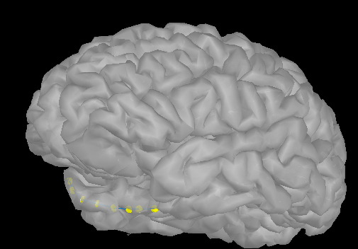
I was then able to right click and select electrode positions from the CT overlay!
We do also use CURRY for this. I guess my goal was to see if Brainstorm could do this independently.
I then chose to resample the CT.
After that I renamed my MRI#2 to CT and then I computed MNI coordinates for the CT.
Then, for the CT I did ‘register to default MRI’
Did you simply try the option "Register" when you imported the CT?
This is what it is supposed to do.
If so, you will face a lot of problems related with memory. Your system may have a lot of RAM in it, but does not allow you to manipulation more than a fraction of it at a time, because of addressing limitations.
If you go to File > Edit preferences, what is the largest variable allowed? (indicated at the bottom-right of this figure)
I have only 8Gb of RAM and I can run these operations without any problem.
Could you send me some example files that I could test? (one MRI + one CT)
Upload them somewhere and them the links to me in a separate email (click on my username on this forum)
Thanks for your example data.
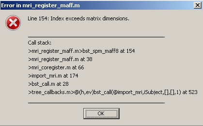
I fixed the two issues: replaced the full interpolation by an interpolation by blocks, and added the step “resample” to the step “register” when needed.
You should be able to do either “resample+register”, or just directly “register”. And it works with less than 6Gb of RAM.
This method (SPM linear registration based on mutual information) aligns beautifully the CT and MR scans you sent me, and takes only about 1min to run!
Thank you for helping me improving these tools.
Francois
FYI: We improved significantly the tools available in Brainstorm for processing and visualizing SEEG and ECOG data, including new options for volume coregistration. They are now documented in a new tutorial: http://neuroimage.usc.edu/brainstorm/Tutorials/Epileptogenicity