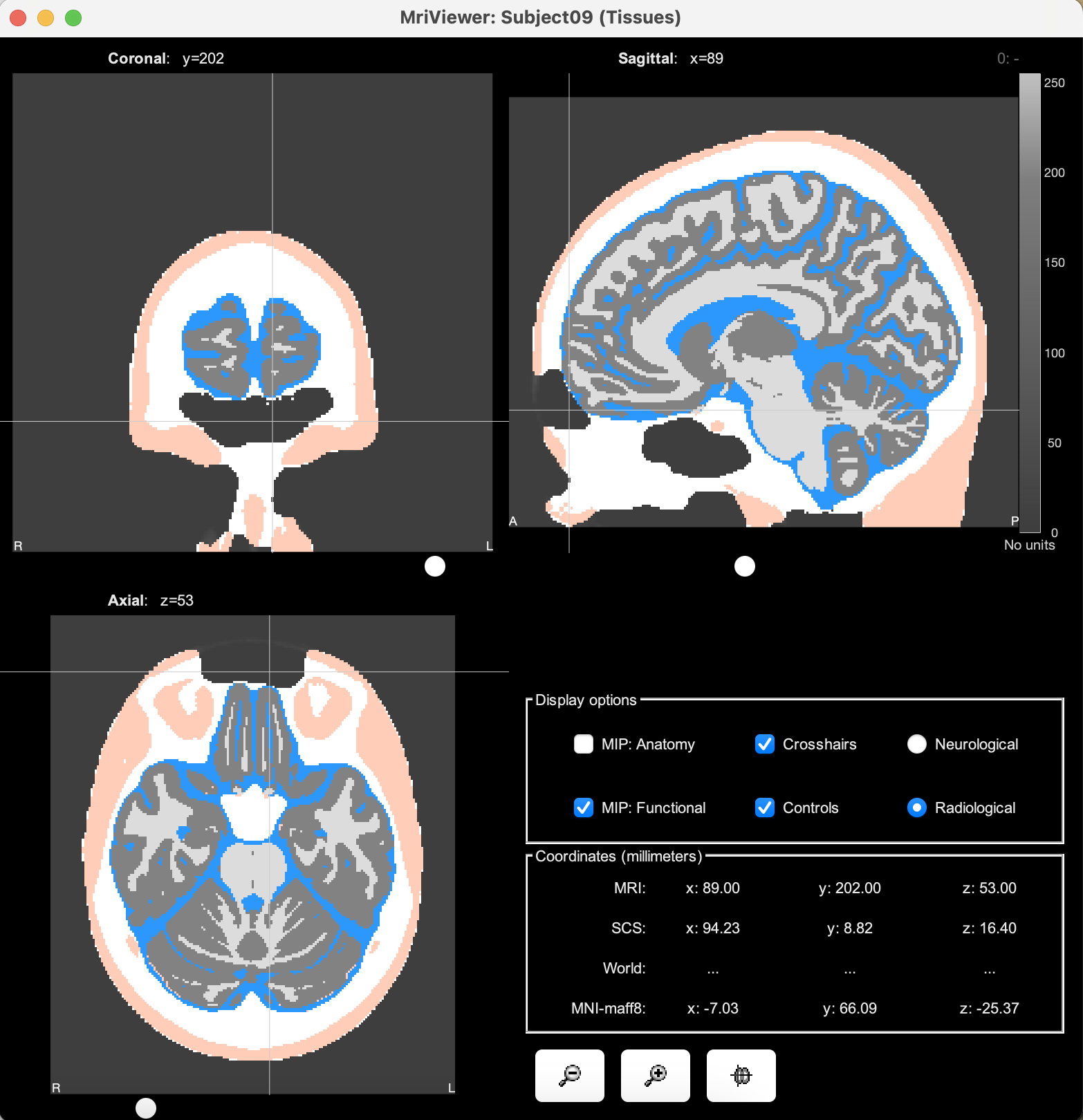
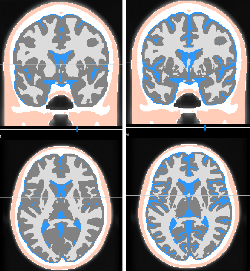
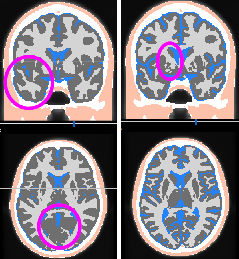
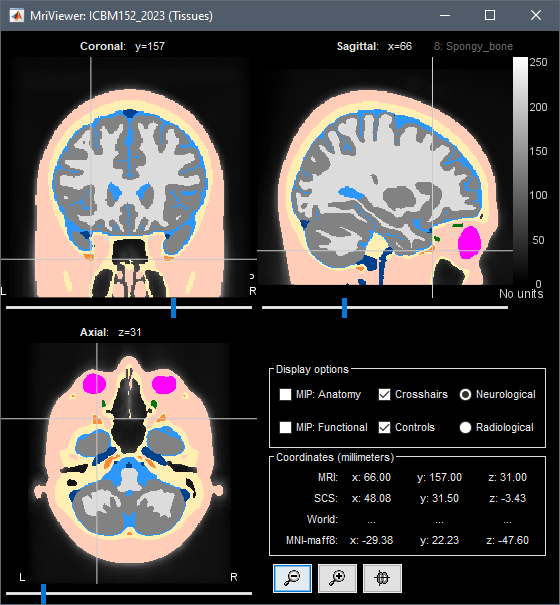
I just tried using the new ICBM template (ICBM 2023) and realized there was something weird with the segmentation in the frontal region (see picture). I am not sure if it's a bug or expected (is it linked to the air in the frontal sinuses ?). Also; it seems that the eye are classified as skin; when i would have thought they would be classified as CSF.
Was this file manually generated by SPM or cat12 or is it directly coming from the template? The description of ICBM indicates that the template includes T1w,T2w,PDw modalities, and tissue probabilities maps.
Also, was the segmentation from FreeSurfer created using only the T1w or the T1w and T2w ?
Oops... this looks pretty bad indeed...
This Tissues file was generated with CAT12 r2166.
The surfaces were generated with FreeSurfer 7.3.2, with the T1 only (this is not a realistic T1 and T2)
I haven't kept track of the decision making for using this sharper and darker volume instead of the old one. Maybe the high contrast and sharp separation between elements was helping with some processes. Or something more irrational, like: I was working on a screen with the brightness set to the max...
Looking again at it now, I agree with you, I don't think it makes much sense, it's exactly the same volume but just darker... I don't even recall where it was coming from...
=> I will restore the older contrast
The FreeSurfer segmentation was computed from the "brighter" volume (the one available on the MNI website), nothing to fix on this end.
The CAT12 segmentation, however, was executed on the dark volume, and this is probably the cause of the problems in segmentation. I'm currently re-running it to get the skin+bone masks, and this time I'll use the csf+wm+gm TPMs from the MNI website.
=> I will update the Tissues volume.
and this time I'll use the csf+wm+gm TPMs from the MNI website.
Now I'm testing this, I'm not sure about it anymore.
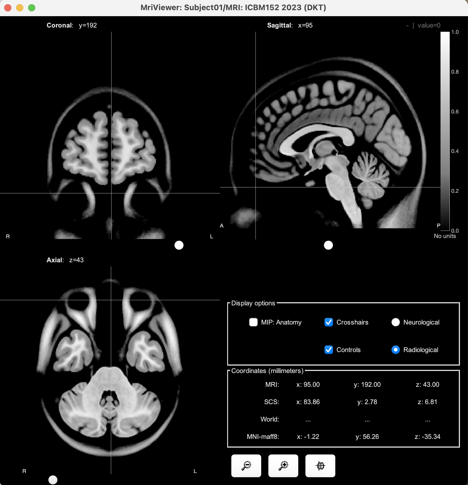
The TPMs from the MNI website (left) produce more realistic voxel classifications than the CAT12 segmentation (right), however the CSF layer is not continuous, which might cause other problems.
oh ok; i see. I thought it was the output of FreeSurfer. It looks indeed better to separate GM and WM. but maybe not great for skin and CSF.
By curiosity how did you combined the output of CAT12 and the one files from the template ? I guess you did something like putting all the voxel where the probability of skin is larger than 0.5 to skin, then Skull, GM then WM ? if yes, maybe instead looking at each vowel which tissue has the largest prob might be better ?
I am asking that since, it seems that the GM is way more large in this segmentation than in the output of CAT12.
By curiosity how did you combined the output of CAT12 and the one files from the template ?
I started from the tissues volume from CAT12, which include all the tissues.
Then I used the volumes from the MNI website to overwrite the values within the brain mask (using the maximum probability at each voxel):
I am asking that since, it seems that the GM is way more large in this segmentation than in the output of CAT12.
Indeed, the GM is much thicker when using the MNI ICBM152 TPMs.
But it might be only detail at the end
In some places there is almost a centimeter of difference for the GM boundaries, so it would have a major impact on any process that uses this tissue classification (example: the automatic labeling of SEEG electrodes).
Is there any simple way to remove/merge specific tissue from the segmentation? i guess i can always export to Matlab and change there but I was wondering if there was a way from Brainstorm GUI?
Also, is there a way to run SimNIBS4/CHARM from Brainstorm?
Is there any simple way to remove/merge specific tissue from the segmentation? i guess i can always export to Matlab and change there but I was wondering if there was a way from Brainstorm GUI?
No, but we need something like this.
I thought however that this is something that would fit better directly in the functions that need the tissues to be merged, so that different pipelines can keep on using the same FEM mesh in the database without having to duplicate it with different combinations of merge tissues (with risks of not selecting the correct file).
How do you use this file, and what would you like to merge and when?
is there a way to run SimNIBS4/CHARM from Brainstorm?