Hello,
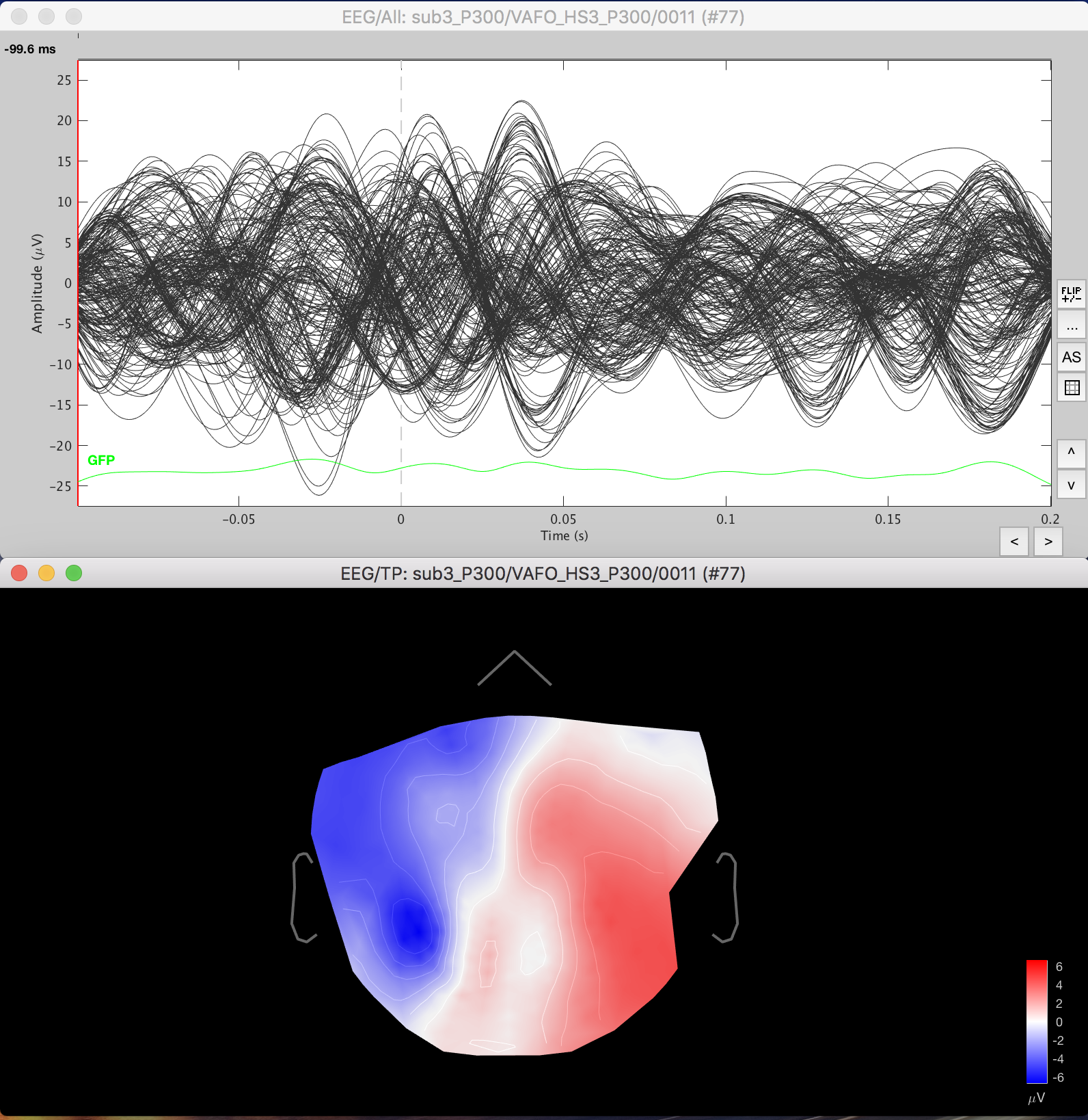
I am working with eeg data recorded with a 256 channel cap (ASA waveguard). When viewing the topography the head model image doesn't correlate correctly to the positions of the electrodes - seems like some areas are missing in temporal and frontal area. However, if I import data from a 64 channel cap the topography appears correct. I tried recorrecting the reference nodes but the topography appears the same.
Is this something that needs updating or something that I can fix within my data?
Thanks!
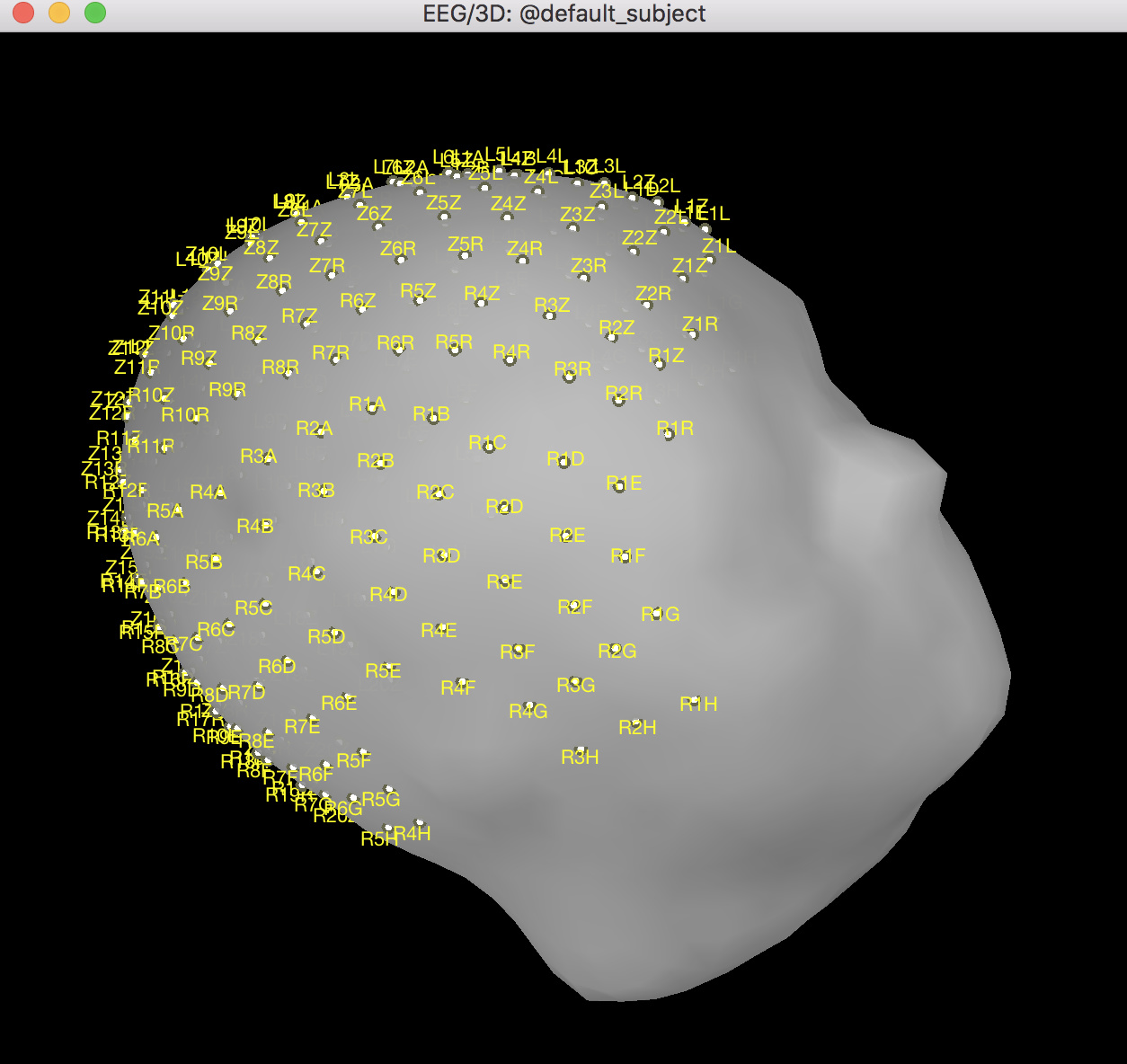
How did you add the 3D positions of the electrodes to your recordings?
Where did you get these 3D positions from?
Please post a screen capture of the 3D positions of the electrodes + the head surface (simply double-click on the channel file should show you that)
I added them by right clicking on the subject and Import channel file.
Do you have any ideas what may cause the weird topographical appearance using the 256 channels?
Rún
How did you get these positions?
From your figure, we don’t see well if they are correctly centered left-right.
This “2D sensor cap” view projects the sensors on a 2D plane, without much supervision, therefore with some headsets, the final shape may look weird.
If you’re not happy with it, you can use the menu “2D Disc” instead.