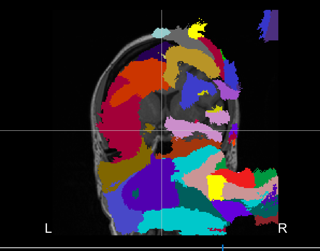
I could reproduce this behavior by running CAT12 from Brainstorm.
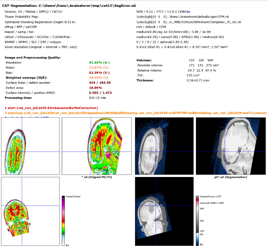
The execution report shows that there is something off with the definition of the volume coordinates (the correspondence between voxels and scanner coordinates).
@tmedani
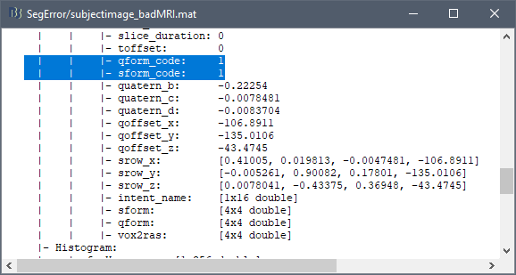
Francois is absolutely right. There is something odd with your SFORM and QFORM matrices. If you visualize the image in SPM12 you see that there is some shearing applied and the voxel sizes are not correct. I have tried to reorient, but there the shearing couldn't be repaired. Please ask for the dicom images of that subject or whether the nifti files are converted using a different tool. This seems to be not nifti-conform even if it looks fine with other tools.
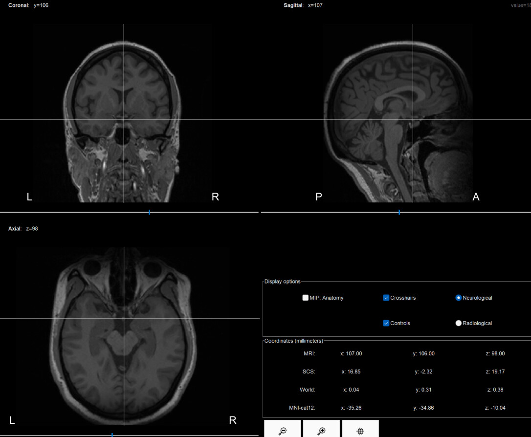
@tmedani We don't see the shearing in Brainstorm, BrainSuite or MRIcron because all these tools display slices of voxels from the cube of values saved in the .nii file. The SFORM/QFORM transformation do not alter the visualization, they only change the computation of the coordinates.
At the contrary, SPM/CAT compute slices through the volume along arbitrary axes (not necessarily aligned with the voxel axes), and including the SFORM or QFORM transformation. Errors in these matrices become visible.
Please ask for the dicom images of that subject or whether the nifti files are converted using a different tool.
I hope the answer is not going to be that the conversion was done with Brainstorm...
This is an update regarding this issue.
I was able to get he original MRIs from the hospital without any processing and the CAT/SPM/SIMNIBS pipelines worked fine.
I want also to highlight that when I used Brainsuite to segment these "BadMRIs", I was able to get good outputs (correct shape and alignement). This mean that Brainsuite get/recompute the missing value from somewhere else at the NiftiFile.
The remaining issue with Brainsuite, in some cases, the WM and GM surface are intersecting, and thus its impossible to generate correct tetramesh with the current tools.