
|
Size: 1211
Comment:
|
Size: 2610
Comment:
|
| Deletions are marked like this. | Additions are marked like this. |
| Line 14: | Line 14: |
| == Dataset description == This dataset includes recordings for a patient that was not reported in the above article, but are part of the same study. The patient presents a focal epilepsy of the left temporo-occipital junction, MRI-negative, and was implanted in the surrounding areas. The subfolder "seeg" contains the recordings of three seizures, all of them showing a propagation of high-frequency oscillations from the lesion towards the temporal lobe, bilaterally. {{attachment:dixi_am.gif||align="right",width="200px"}} The depth electrodes used in this example dataset are [[http://www.diximedical.com/en/microdeep-depth-electrode|DIXI D08-**AM Microdeep electrodes]], with the following specifications: * Diameter: 0.8 mm * Contact length: 2 mm * Insulator length: 1.5 mm * Distance between the center of two contacts: 3.5 mm * Between 8 and 18 contacts on each electrode Files included in this package: * anat/MRI/3DT1pre_deface.nii: Subject MRI before SEEG implantation * anat/MRI/3DT1post_deface.nii: Subject MRI after SEEG implantation * anat/MRI/brainvisa: Cortical surface extracted with BrainVISA 4.5 * anat/implantation/*: Positions of the SEEG contacts in various formats (MNI or subject space) * seeg/SZ*.TRC: Seizure recordings in Micromed format, one seizure per file * MRI scans were de-identified with FreeSurfer's mri_deface. |
SEEG/Epileptogenicity maps
License
This tutorial dataset (EEG and MRI data) remains property of the Grenoble University Hospital, France. Its use and transfer outside the ImaGIN tutorial, e.g. for research purposes, is prohibited without written consent. For questions, please contact Olivier David, PhD ( olivier.david@grenoble-univ-alpes.fr ).
Reference
The acquisition methodology is described in the following articles:
David O, Blauwblomme T, Job AS, Chabardès S, Hoffmann D, Minotti L, Kahane P, Imaging the seizure onset zone with stereo-electroencephalography, Brain. 2011 Oct;134(10):2898-911
Lamarche F, Job AS, Deman P, Bhattacharjee M, Hoffmann D, Gallazzini-Crépin C, Bouvard S, Minotti L, Kahane P, David O, Correlation of FDG-PET hypometabolism and SEEG epileptogenicity mapping in patients with drug-resistant focal epilepsy, Epilepsia. 2016 Dec; 57(12):2045–2055
Dataset description
This dataset includes recordings for a patient that was not reported in the above article, but are part of the same study. The patient presents a focal epilepsy of the left temporo-occipital junction, MRI-negative, and was implanted in the surrounding areas. The subfolder "seeg" contains the recordings of three seizures, all of them showing a propagation of high-frequency oscillations from the lesion towards the temporal lobe, bilaterally.
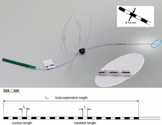
The depth electrodes used in this example dataset are DIXI D08-**AM Microdeep electrodes, with the following specifications:
- Diameter: 0.8 mm
- Contact length: 2 mm
- Insulator length: 1.5 mm
- Distance between the center of two contacts: 3.5 mm
- Between 8 and 18 contacts on each electrode
Files included in this package:
- anat/MRI/3DT1pre_deface.nii: Subject MRI before SEEG implantation
- anat/MRI/3DT1post_deface.nii: Subject MRI after SEEG implantation
- anat/MRI/brainvisa: Cortical surface extracted with BrainVISA 4.5
- anat/implantation/*: Positions of the SEEG contacts in various formats (MNI or subject space)
- seeg/SZ*.TRC: Seizure recordings in Micromed format, one seizure per file
MRI scans were de-identified with FreeSurfer's mri_deface.