|
Size: 191
Comment:
|
Size: 1807
Comment:
|
| Deletions are marked like this. | Additions are marked like this. |
| Line 4: | Line 4: |
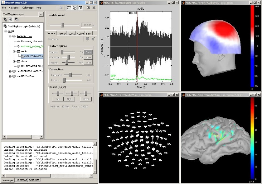
| == MEG Auditory evoked potential == Acquisition system: ''Neuromag Vectorview306''. Description of the windows: * Main !BrainStorm window * Timeseries of all the MEG sensors [-200ms, 500ms] * Magnetic field recorded by the magnetometers at t=106ms * Spatial view of the magnetometers time series [-200ms, 500ms] * Reconstruction of the cortical currents, based on the magnetometers, at t=106ms [[attachment:snap_1condition.jpg|{{attachment:snap_1condition_sm.jpg|attachment:snap_1condition.jpg}}]] |
|
| Line 5: | Line 18: |
| ||<tablewidth="100%"><style="border:0px;"> {{attachment:snap_1condition.jpg||height="353",width="504"}} ||<style="vertical-align: top;">Standard view || | |
| Line 7: | Line 19: |
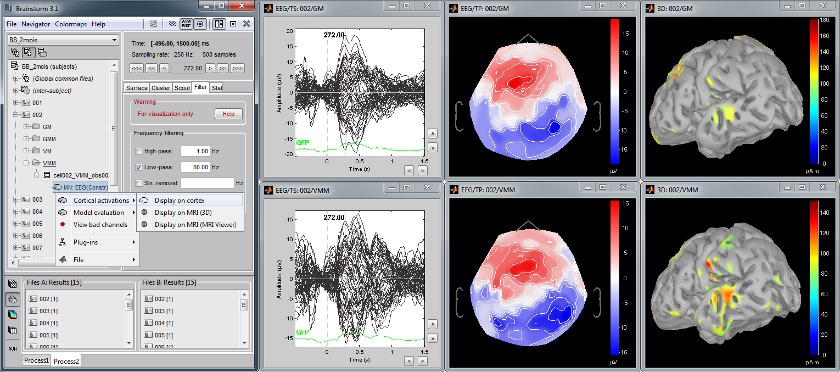
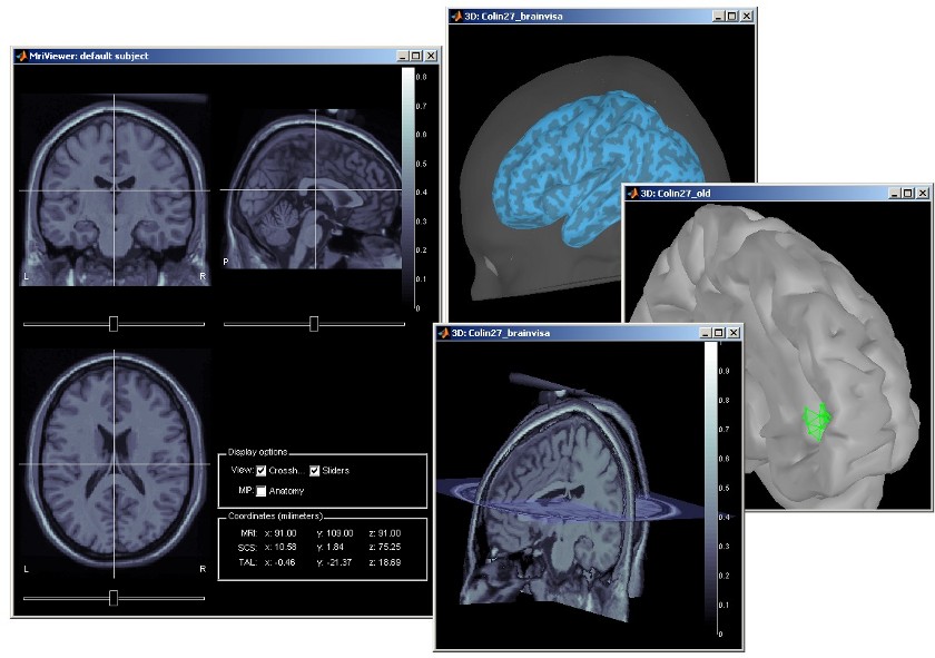
| == Multiple conditions: Baby auditory EEG == Acquisition system: ''EGI GSN - 64 electrodes'' Description: * One subject: "001" * Three conditions: "GM", "GMM", "VM" * Two views: overlaid electrodes time series, and estimated cortical sources at t=376ms [[attachment:snap_3conditions.jpg|{{attachment:snap_3conditions_sm.jpg|attachment:snap_3conditions.jpg}}]] <<BR>> == Subject anatomy: MRI and surfaces == BrainStorm offers the possibility to reconstruct the cortical activity either on the individual subject anatomy, or on a default anatomy (MNI / Colin27). For this purpose, many interactive tools are available to view, register and process the MR images and the corresponding meshes. However, the segmentation must be performed by external program of your choice ([[Links|list here]]). Here are a few examples of the views you can obtain with a simple click on the subject's MRI or surface. All the 3D views can be rotated freely with the mouse, zoomed with the wheel, edited with the "Surfaces panel" and with their popup menu. The MRI slices can be moved with a simple mouse operation: right-click and mouse drag. [[attachment:snap_anatomy.jpg|{{attachment:snap_anatomy_sm.jpg|attachment:snap_anatomy.jpg}}]] |
Screenshots
MEG Auditory evoked potential
Acquisition system: Neuromag Vectorview306.
Description of the windows:
Main BrainStorm window
- Timeseries of all the MEG sensors [-200ms, 500ms]
- Magnetic field recorded by the magnetometers at t=106ms
- Spatial view of the magnetometers time series [-200ms, 500ms]
- Reconstruction of the cortical currents, based on the magnetometers, at t=106ms
Multiple conditions: Baby auditory EEG
Acquisition system: EGI GSN - 64 electrodes
Description:
- One subject: "001"
- Three conditions: "GM", "GMM", "VM"
- Two views: overlaid electrodes time series, and estimated cortical sources at t=376ms
Subject anatomy: MRI and surfaces
BrainStorm offers the possibility to reconstruct the cortical activity either on the individual subject anatomy, or on a default anatomy (MNI / Colin27). For this purpose, many interactive tools are available to view, register and process the MR images and the corresponding meshes. However, the segmentation must be performed by external program of your choice (?list here).
Here are a few examples of the views you can obtain with a simple click on the subject's MRI or surface. All the 3D views can be rotated freely with the mouse, zoomed with the wheel, edited with the "Surfaces panel" and with their popup menu. The MRI slices can be moved with a simple mouse operation: right-click and mouse drag.


