|
Size: 549
Comment:
|
Size: 5203
Comment:
|
| Deletions are marked like this. | Additions are marked like this. |
| Line 1: | Line 1: |
| = Screenshots = <<BR>> |
= Gallery = == Demo video == Tadel F & Baillet S, March 2015<<BR>>Brainstorm: Imaging neural activity at the speed of brain ([[http://neuroimage.usc.edu/videos/201503_brainstorm_demo.mp4|Download video]]) |
| Line 4: | Line 5: |
| <<BR>> || {{attachment:snap_1condition.jpg||height="373",width="534"}} ||<style="vertical-align: top;">MEG auditory evoked potential<<BR>> (Neuromag Vectorview306)<<BR>> Description of the windows (left to right): * Main BrainStorm window * Timeseries of all the MEG sensors [-200ms, 500ms] * Magnetic field recorded by the magnetometers at t=106ms * Spatial view of the magnetometers time series [-200ms, 500ms] * Reconstruction of the cortical currents, based on the magnetometers, at t=106ms || |
<<HTML(<iframe id="ytplayer" type="text/html" width="853" height="480" src="http://www.youtube.com/embed/30eFJUrRcN4?autoplay=1&origin=http://http://neuroimage.usc.edu/brainstorm" frameborder="0"/>)>> |
| Line 7: | Line 7: |
| <<HTML(<video width="853" height="480" controls><source src="http://neuroimage.usc.edu/videos/201503_brainstorm_demo.mp4" type="video/mp4"><source src="http://neuroimage.usc.edu/videos/201503_brainstorm_demo.ogg" type="video/ogg">Your browser does not support the video tag.</video>)>> | |
| Line 8: | Line 9: |
| == MEG somatosensory evoked responses == Acquisition on a ''CTF 275'' instrument for a left median nerve electric stimulation. <<BR>>[[attachment:snap_median.jpg|{{attachment:snap_median_sm.jpg|attachment:snap_median.jpg}}]] |
|
| Line 9: | Line 12: |
| == Baby auditory EEG responses == Acquisition system: ''EGI GSN - Baby 64 electrodes''. <<BR>> [[attachment:snap_3conditions.jpg|{{attachment:snap_3conditions_sm.jpg|attachment:snap_3conditions.jpg}}]] |
|
| Line 10: | Line 15: |
| ---- <<BR>> |
== Continuous recordings and markers == Review recordings directly reading from the original files, edit markers, detect and correct artifacts. <<BR>> [[attachment:snap_raw.jpg|{{attachment:snap_raw_sm.jpg|attachment:snap_raw.jpg}}]] == Artifact detection and correction with SSP == A fully automated cleaning pipeline for the most common artifacts (power lines, eye blinks, heartbeats) is available in Brainstorm. The ocular and cardiac artifacts are corrected using the [[Tutorials/TutRawSsp|Signal Space Projection]] approach. === Example: Heartbeat === {{attachment:ssp_ecg.jpg}} === Example: Eye blink === [[attachment:sspExample.gif|{{attachment:sspExample_sm.jpg|attachment:sspExample.gif}}]] == Subject anatomy: MRI and surfaces == Brainstorm features the possibility to model MEG and EEG neural generators either from the individual subject anatomy, or by using a template anatomy (MNI / Colin27) that can be warped to the individual scalp surface. Multiple interactive tools are available to view, register and process the MR images and the corresponding tessellated envelopes. However, tissue segmentation must be performed using another software; multiple options exist today in the academic community ([[Links|listed here]]). We provide a few examples of the views you can easily obtain with Brainstorm. All the 3D views can be rotated freely with the mouse, zoomed with the wheel, edited with the "Surface panel" and contextual popup menus. The MRI slices can be browsed with a simple mouse operation: right-click and mouse drag.<<BR>>[[attachment:snap_anatomy.jpg|{{attachment:snap_anatomy_sm.jpg|attachment:snap_anatomy.jpg}}]] == Channel selection == All the figures displayed by Brainstorm are linked in time. If they feature the same dataset, the sensor selection is also the same for all views. The selection of a channel subset can be easily perfomed by clicking on the corresponding channels in a time series display or a 3D view. Selected channels can be displayed separately, marked as "bad", or deleted.<<BR>>[[attachment:snap_channel.jpg|{{attachment:snap_channel_sm.jpg|attachment:snap_channel.jpg}}]] == Cortical regions of interest == Acquisition system: ''CTF MEG - 275 sensors'' Scouts are cortical regions of interest, defined graphically from the "Scout" tab. They can be used to extract the time series of MEG and EEG generators within a single or mulitlple brain region. The following example shows the cortical response to an electric stimulation of the left median nerve. With the three scouts "S1 right", "S2 right" and "S2 left"'','' we can track the processing of the stimulus in the brain: 1) contralateral primary somatosensory cortex, 2) contralateral sencondary somatosensory, 3) ipsilateral secondary somatosensory.<<BR>>[[attachment:snap_1scout.jpg|{{attachment:snap_1scout_sm.jpg|attachment:snap_1scout.jpg}}]] == Anatomical atlases == Integrated support for the individual surface-based anatomical atlases generated by FreeSurfer and BrainSuite.<<BR>><<BR>> {{attachment:atlases.jpg||height="358",width="750"}} == Scripting environment == Everything that can be done in the interface with mouse clicks can be converted automatically to Matlab scripts, using the Process1 and Process2 tabs.<<BR>>[[attachment:snap_scripting.jpg|{{attachment:snap_scripting_sm.jpg|attachment:snap_scripting.jpg}}]] == Digitize and co-register == For accurate source localization in MEG and EEG with registration to anatomical MRI image volume and efficient head localization in MEG, a 3D digitization solution such as the Polhemus-Fastrak is required. Brainstorm features an integrated support for driving a Polhemus device that will help you save time and is efficient for error-checking while digitizing sensor positions and the subject's head shape.<<BR>>The digitized points are used to check and/or fix visually the alignment of the MRI/surfaces and the MEG/EEG sensors.<<BR>><<BR>> {{attachment:polhemus.jpg}} == Functional connectivity == {{attachment:connect_graph.jpg}} |
Gallery
Demo video
Tadel F & Baillet S, March 2015
Brainstorm: Imaging neural activity at the speed of brain (Download video)
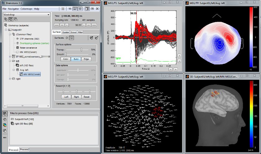
MEG somatosensory evoked responses
Acquisition on a CTF 275 instrument for a left median nerve electric stimulation.
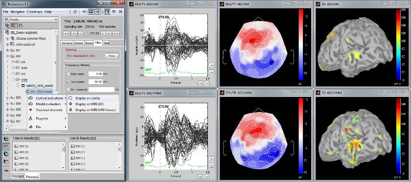
Baby auditory EEG responses
Acquisition system: EGI GSN - Baby 64 electrodes.
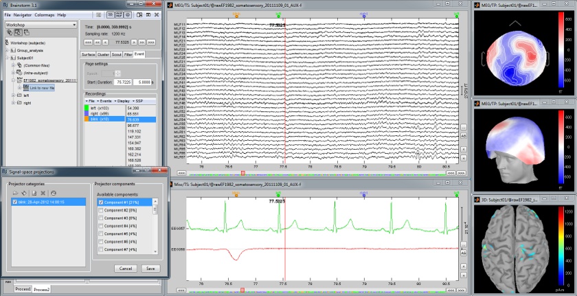
Continuous recordings and markers
Review recordings directly reading from the original files, edit markers, detect and correct artifacts.
Artifact detection and correction with SSP
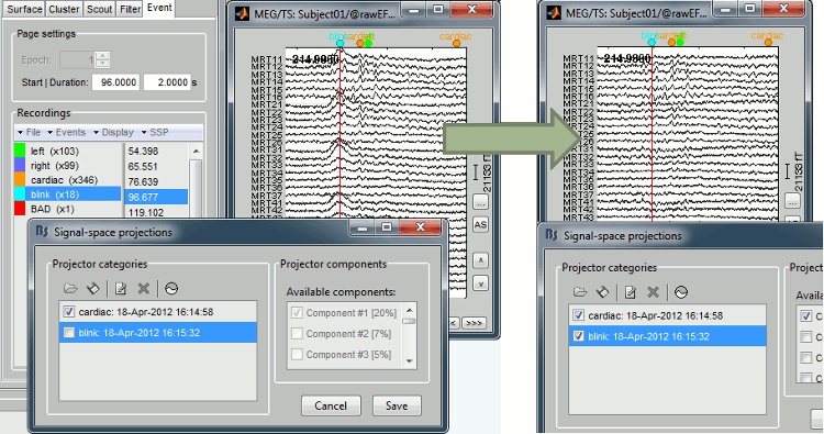
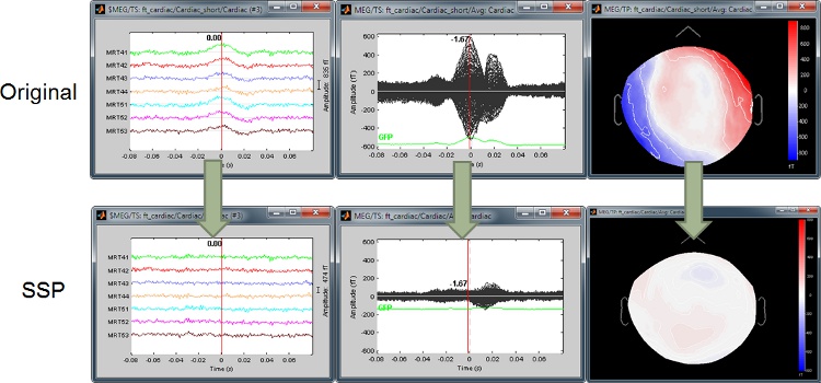
A fully automated cleaning pipeline for the most common artifacts (power lines, eye blinks, heartbeats) is available in Brainstorm. The ocular and cardiac artifacts are corrected using the ?Signal Space Projection approach.
Example: Heartbeat
Example: Eye blink
Subject anatomy: MRI and surfaces
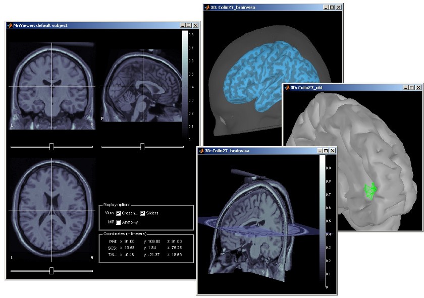
Brainstorm features the possibility to model MEG and EEG neural generators either from the individual subject anatomy, or by using a template anatomy (MNI / Colin27) that can be warped to the individual scalp surface. Multiple interactive tools are available to view, register and process the MR images and the corresponding tessellated envelopes. However, tissue segmentation must be performed using another software; multiple options exist today in the academic community (?listed here).
We provide a few examples of the views you can easily obtain with Brainstorm. All the 3D views can be rotated freely with the mouse, zoomed with the wheel, edited with the "Surface panel" and contextual popup menus. The MRI slices can be browsed with a simple mouse operation: right-click and mouse drag.
Channel selection
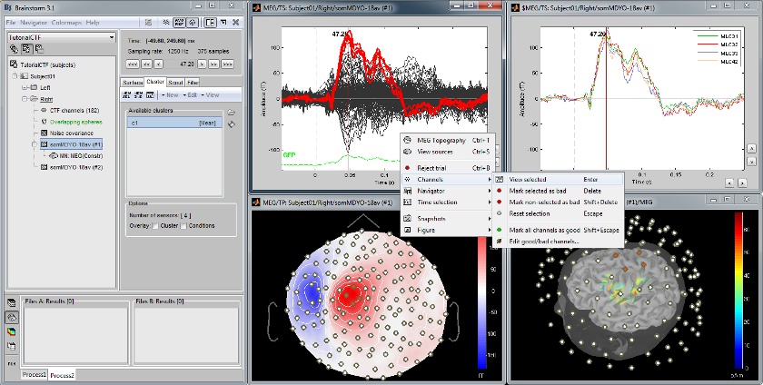
All the figures displayed by Brainstorm are linked in time. If they feature the same dataset, the sensor selection is also the same for all views. The selection of a channel subset can be easily perfomed by clicking on the corresponding channels in a time series display or a 3D view. Selected channels can be displayed separately, marked as "bad", or deleted.
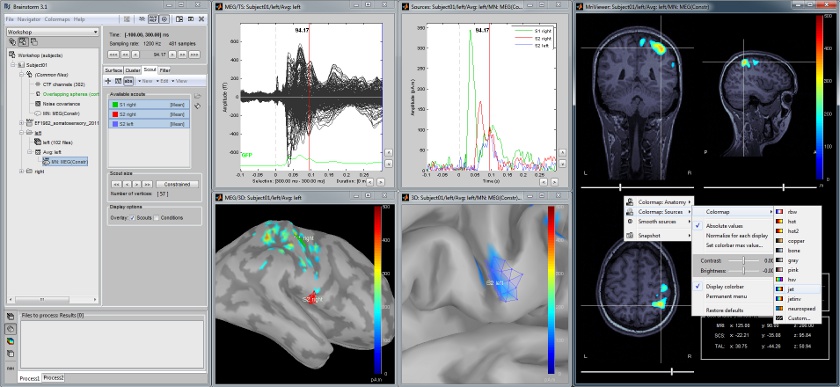
Cortical regions of interest
Acquisition system: CTF MEG - 275 sensors
Scouts are cortical regions of interest, defined graphically from the "Scout" tab. They can be used to extract the time series of MEG and EEG generators within a single or mulitlple brain region.
The following example shows the cortical response to an electric stimulation of the left median nerve. With the three scouts "S1 right", "S2 right" and "S2 left", we can track the processing of the stimulus in the brain: 1) contralateral primary somatosensory cortex, 2) contralateral sencondary somatosensory, 3) ipsilateral secondary somatosensory.
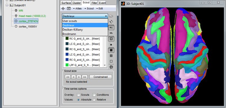
Anatomical atlases
Integrated support for the individual surface-based anatomical atlases generated by FreeSurfer and BrainSuite.
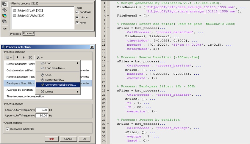
Scripting environment
Everything that can be done in the interface with mouse clicks can be converted automatically to Matlab scripts, using the Process1 and Process2 tabs.
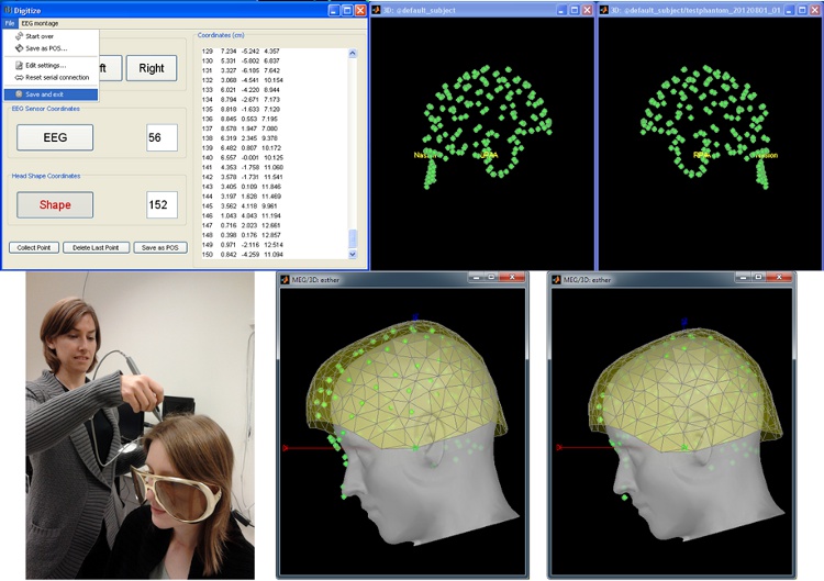
Digitize and co-register
For accurate source localization in MEG and EEG with registration to anatomical MRI image volume and efficient head localization in MEG, a 3D digitization solution such as the Polhemus-Fastrak is required. Brainstorm features an integrated support for driving a Polhemus device that will help you save time and is efficient for error-checking while digitizing sensor positions and the subject's head shape.
The digitized points are used to check and/or fix visually the alignment of the MRI/surfaces and the MEG/EEG sensors.
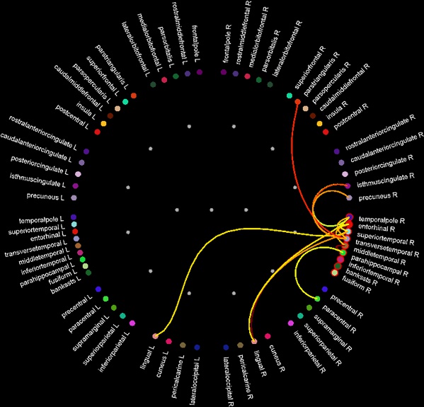
Functional connectivity
