A few problems with mixed models

Hi Martin/Francois,

I am working with mixed models for the first time in a while and have noticed a few curiosities. (Brainstorm is updated to 13 April 2020; Matlab 2019a)

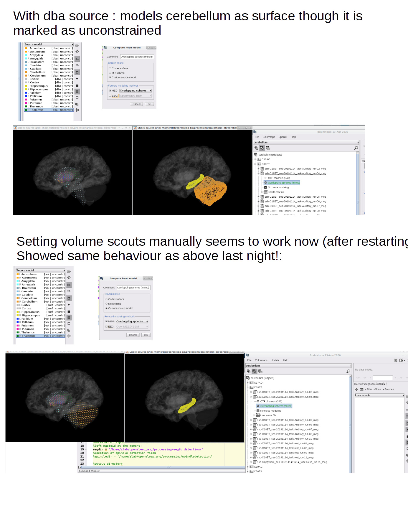
1. Forward mixed model assigns cerebellum to surface rather than volume, and not cortex.
Steps:

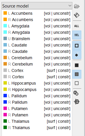
  • under Anatomy tab, I create a source model option atlas will all structures (which include cortex and subcortical regions) set to deep brain (dba). The structures correctly default to cortex as surface (constr), subcortical structures as volume (unconstrained).
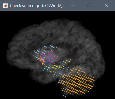
  • under the Functional tab, I create a mixed overlapping spheres head model. Looking at the grid (Check source grid (volume) ), the cerebellum is not included in the volume portion, though the other non-cortical structures are. Looking at the surfaces, only hippocampus and cerebellum are represented (not pallidum and not cortex)
  • Troubleshooting attempt: if I go back to the anatomy tab Source Model atlas and set everything to Volume, except for cortex and thalamus to surface, and repeat the overlapping spheres head model, it does not correct the problem - cerebellum still appears to be a surface and cortex doesn't exist at all, from which I conclude it is not the dba defaults but rather something more general.

2. Forward mixed model calculation allows you to set different grid sizes, but ignores the argument.
Steps:

  • for mixed models, the option to change grid size can be accessed if you run the forward model via the Process 1 tab. (Compute head model > Custom source model, MRI volume grid: Edit: Regular grid (isotropic), grid resolution ....)
  • however, changing the value doesn't change the outcome, which seems to default to a 5mm iso grid regardless of the numbers you put in. (This grid size is inadequate for some of the smaller structures)
  • Troubleshooting attempt: I redid the overlapping spheres head model with the Generate from Cortical Surface option and visualized it, and it still looks like a 5mm iso grid only on the brainstem and subcortical regions.

3. Importing volume masks on mixed models (I don't know if this is a bug or just not possible yet)

  • I have a subject-space subcortical volume-based mask that works nicely on full-volume models. If I read it into a mixed model, it doesn't give an error and adds an atlas with scouts, but I don't see anything appear on the brain visualization.
  • Troubleshooting attempt: I tried to read it in on a full-volume model, export the scouts to matlab, and read them in on the mixed model that had the same iso grid. They appear, but not in the right place. Is there any way to read in subcortical masks on mixed models? (That would be very useful!)

Best regards,

Emily

Forward mixed model assigns cerebellum to surface rather than volume

This is what the authors of this method recommended. According to them the orientation of the sources in the cerebellum is mainly orthogonal to its surface.
https://neuroimage.usc.edu/brainstorm/Tutorials/DeepAtlas#References

This parameter is highly questionable in the context of Brainstorm, because we don't have access to an accurate surface of the cerebellum with its complex folding. I would tend to think that either cortical/unconstrained or volume source model is more appropriate for the cerebellum.

Troubleshooting attempt: if I go back to the anatomy tab Source Model atlas and set everything to Volume, except for cortex and thalamus to surface, and repeat the overlapping spheres head model, it does not correct the problem - cerebellum still appears to be a surface and cortex doesn't exist at all, from which I conclude it is not the dba defaults but rather something more general.

I can't reproduce this behavior. I used the Brainstorm introduction tutorials, created a mixed source model with all the subcortical structures from the ASEG atlas, assign everything to volume except cortex and thalamus, and got what I asked for:
image image
Make sure you (or Brainstorm) didn't get confused with multiple "cortex_mixed" surfaces in the subject anatomy.

Forward mixed model calculation allows you to set different grid sizes, but ignores the argument.

In the process "Compute head volume", the parameter "MRI volume grid" is only related with the option "Source space: MRI volume".
In the case of "Source space: Custom source model", the definition of the all the grids of sources (surface and volume) is handled by the functions of the DBA toolbox:
https://github.com/brainstorm-tools/brainstorm3/blob/master/external/dba/dba_anatmodel.m#L13

If you want to set the grid resolution, simply use the option "MRI volume" instead. It might be a better option in general... or at least it makes the results much easier to process.

Importing volume masks on mixed models (I don't know if this is a bug or just not possible yet)

Not possible, sorry...
Interactions between scouts and mixed models are complicated, and this is not really our priority to improve these models at the moment.
Do you really think this improves the interpretation of the source localization compared with either a simpler surface or volume source model?

If you're curious about improvements on source modeling accuracy in Brainstorm, we are working on using realistic FEM models (not ready yet, but probably fully available and tested before the summer):
https://neuroimage.usc.edu/brainstorm/Duneuro
https://neuroimage.usc.edu/brainstorm/meshGeneration

Hi Francois,

Thanks for the response. I agree that our anatomy isn't generally good enough to do accurate cerebellar surface models. (We can partly get around it by modelling the cerebellum as a volume and chopping it into reasonable ROIs that mimic the folded organization of the structure, which seems to be working).

Yesterday evening I was having the described problem with any combination of inputs. Today (after restarting everything) it seems to only be acting oddly using the deep brain default options, see below.

In general the mixed models are harder to work with and have fewer options, so I might abandon them for the current project. In general we use them because it is hard to reconstitute a reasonable-looking timecourse from the unconstrained models (which is necessary for detecting some events, to show equivalence with brain signals that are well known, and I'm not totally convinced connectivity analyses work well with the 3 dimensions), and Sylvain has continued to recommend them for our auditory questions that involve both cortical and subcortical ROIs.

I'm curious to try the new models (and understand why/when they are preferred). In the meantime we are doing some work to compare model parameters for deep sources with the existing options.

Best,

E

Yesterday evening I was having the described problem with any combination of inputs. Today (after restarting everything) it seems to only be acting oddly using the deep brain default options, see below.

I'm not sure to understand what is incorrect in the screen capture you posted.
Do you think there is something to fix?

I'm not totally convinced connectivity analyses work well with the 3 dimensions

Indeed, they don't. And we don't have any statistical tests to suggest either. The question of processing unconstrained sources has been suspended for years...
@Sylvain @John_Mosher @pantazis @hossein27en @leahy Any update on this front?

Sylvain has continued to recommend them for our auditory questions that involve both cortical and subcortical ROIs

Then follow his recommendations.

Good luck with working with all these incomplete options! :slight_smile:

Hi Francois,

I believe that when the Source model option for the cerebellum are set to "unconstrained" and we create an overlapping spheres head model, the cerebellum should become a volume rather than a surface (top set of images). Is that not correct? I had deleted all the head models prior to redoing this, so no chance of a mix up.

Best,

Emily

Following other apps in the field, a simple approach consists in extracting the first principle component - i.e. perform a PCA - from each location in the volume source model. The resulting restricted model would be similar to and could be processed as a surface source model: one time series per source location.

This can be done with process "Sources > Unconstrained to flat map", but the default for connectivity and statistics is still to use the norm instead of the PCA. And there is no mention of this option in the tutorials:
https://neuroimage.usc.edu/brainstorm/Tutorials/Workflows#Unconstrained_cortical_sources

@Sylvain Would you like to propose this to the rest of the group during tomorrow's meeting?

1 Like

Hi Sylvain. Yes, we're doing that, and will try your suggestion about checking the first few components. I'm not sure how to test how well it works for something like connectivity analysis. We have results but are not yet sure if they are sensible. We're continuing to explore.

The problem we're running into for one of the sleep projects is that we want to use subject-space volume ROIs, which can't currently be read in on the mixed models. I the past I avoided the problem (for the auditory projects) by using surface ROIs from one of the freesurfer atlases (which works) and then adding subcortical ROIs based on growing small scouts from seed points based on MNI coordinates, using the MNI-to-subject transformation. That works for roughly spherical regions but now I have more complicated thalamic regions defined by DWI I think we're stuck using the full volume models. (Do you know of anyone who's used thalamic surface models? I don't know if that's reasonable.)

Thanks,

Emily

No, if you create set it to dba/unconstrained, it uses the default option of the DBA method (cerebellum=>surface), resulting in a source model with constrained locations (cerebellum surface) and unconstrained orientations. Just like when you compute regular surface-based forward models with the unconstrained option.
If you want a volume, you have to select "volume" instead of "dba".

We used a crude surface approximation in https://www.ncbi.nlm.nih.gov/pmc/articles/PMC6856176/

1 Like

Aah, okay thanks for the clarification.