Even though I have already used CAT12 and Brainsuite for brain segmentation, they can't provide me with an atlas (like AAL or Diedrichsen2009) registered on the 3D cortex view of the cerebellum. CAT12 can segment the cerebellum using AAL atlas but only in the MRI view (3D or MRI viewer).
Is there a way to register an atlas like AAL in the 3D cortex view of the cerebellum so I can finally use it for the source localization?
Kind regards,
Konstantinos Tsilimparis
Research Associate at Medical Physics Lab
Aristotle University of Thessaloniki
CAT12 cannot be used reliably for the cerebellum surface computation for the moment.
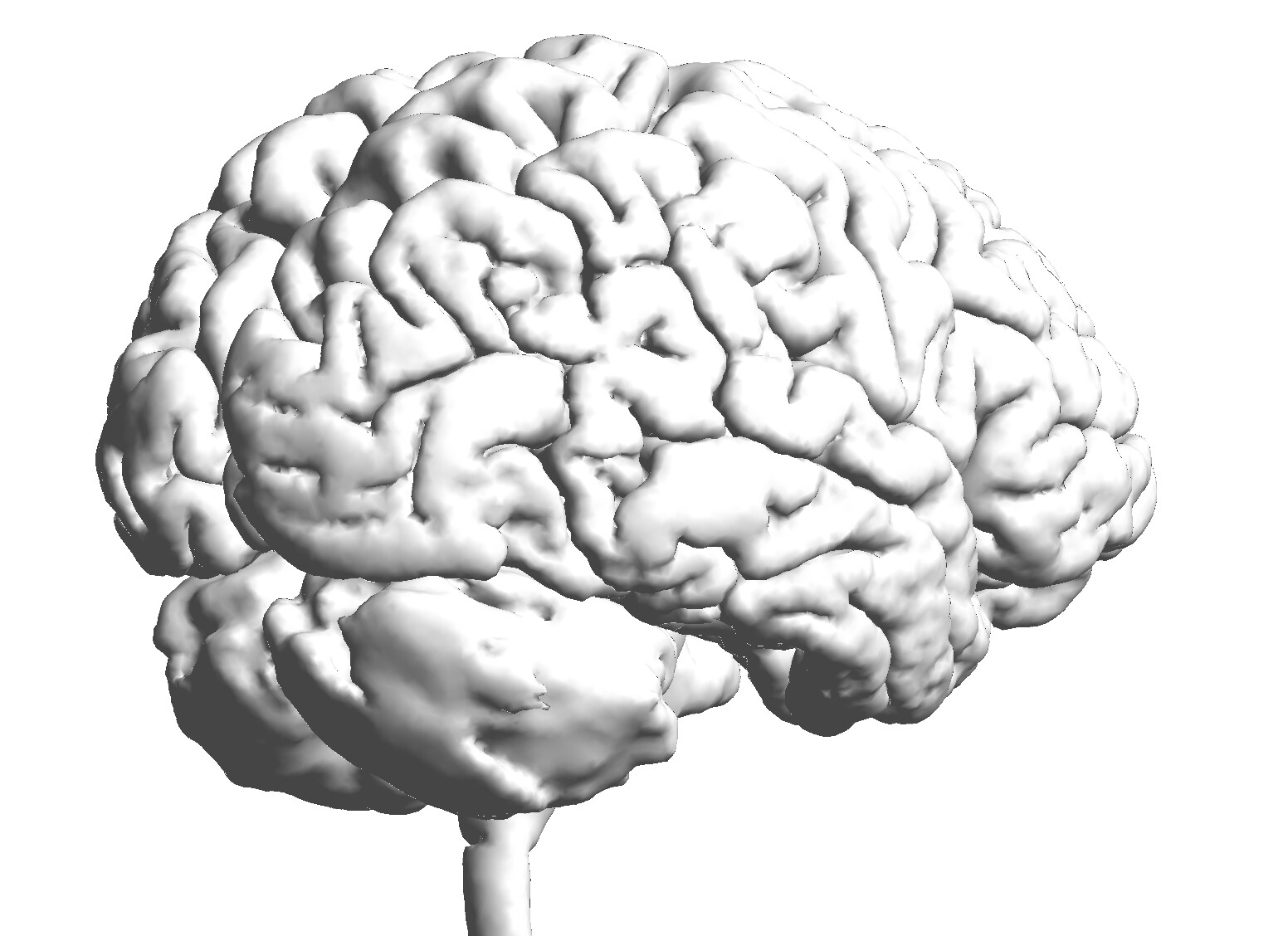
What Brainstorm does to create this surface is simply compute an isosurface of the cerebellum label of the ASEG atlas from FreeSurfer. This provides a correct shape, but not a proper reconstruction of the surface of the cerebellum, and with no registration to any atlas.
Is there a way to register an atlas like AAL in the 3D cortex view of the cerebellum so I can finally use it for the source localization?
AAL is not available on the surface, it is a volume parcellation in MNI-space that is projected into the subject space using non-linear MNI normalization of the subject T1 volume with SPM or CAT.
There is no solution to obtain an AAL parcellation of the surface.
My question is: How does brainstorm take a volume parcellation atlas (.nii file) and projects it on the surface? Is it a reliable method?
Also, when I import the Buckner17 Atlas, brainstorm splits the scouts in right and left direction. For example scout 2 is split into scout 2 R and 2 L. How can I neglect this split and keep the original atlas' scouts?
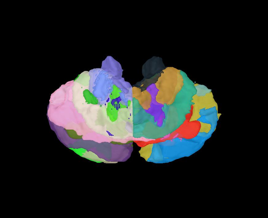
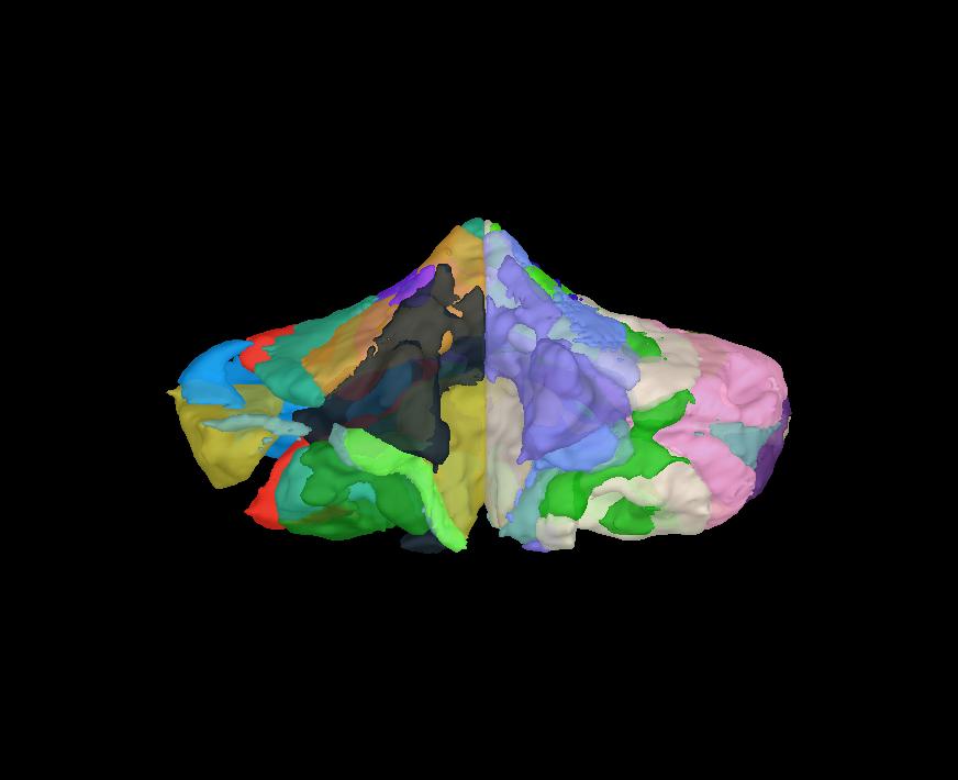
Here are pictures of the cerebellum surface parcellation with Buckner17 in brainstorm:
I import the surface file (right-click on subject > Import surface > Select: atl-Buckner17_space-MNI_dseg.nii).
My question is: How does brainstorm take a volume parcellation atlas (.nii file) and projects it on the surface? Is it a reliable method?
There is no "Projection on the surface".
This menu "Import surface" applied to a volume parcellation creates a new surface file which includes the tesselation of each parcel (computed with Matlab isosurface function).
The output of such procedure, beyond visualization, is very limited.
Also, when I import the Buckner17 Atlas, brainstorm splits the scouts in right and left direction. For example scout 2 is split into scout 2 R and 2 L. How can I neglect this split and keep the original atlas' scouts?
What you get here are not really scouts, but actual surfaces... it does not make much sense to merge them.
If you want to use the parcels from your atlas as scouts in the context of a source analysis, then the best solution is maybe to use a volume source model and load the atlas a volume atlas: https://neuroimage.usc.edu/brainstorm/Tutorials/TutVolSource#Volume_atlases
Sorry for bring this topic uplist again but, I was wondering:
Is there any way I can segment the brain (from DICOM file) and the output surfaces include the cerebellum?
How can I include the subject's cerebellum in the source analysis?
thanks ind advance!
PS: cerebellum segmentation is still unavailable on CAT software?
PS: cerebellum segmentation is still unavailable on CAT software?
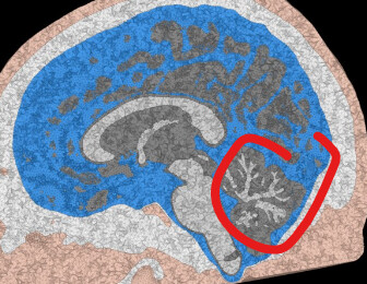
I believe it's now available. Here is an example of the output from the simnibs when cat is used
@tmedani@tourette95
Surface extraction of the cerebellum is still experimental and not recommended. The resulting quality is simply too poor because of the many foldings.
However there is a cerebellar atlas available from the SUIT toolbox by Diedrichsen in newer CAT12 version for volumes.