I want to display the ECoG signal on the macaque cortex. (using the anatomical dataset in the tutorial).
* I use Brainstorm standalone on Windows 10.
First, MRI data were aligned and surface data were imported according to the procedure manual.
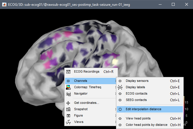
Then, I imported my ECoG data and displayed it on the cortex with "ECoG -> display on cortex".
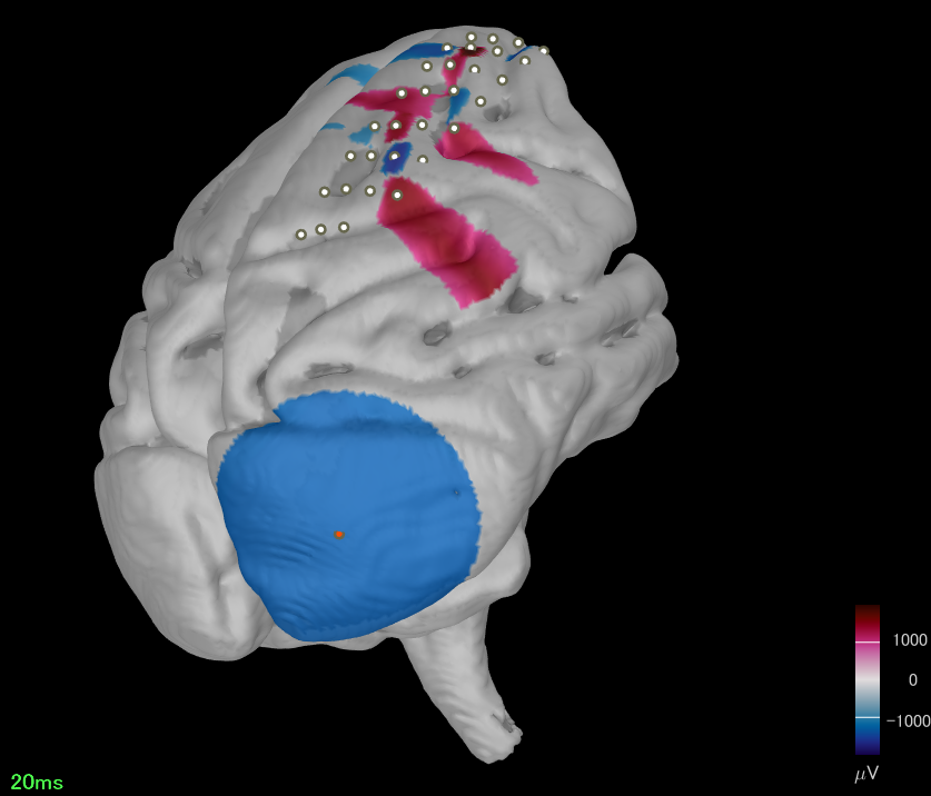
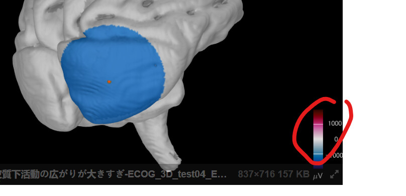
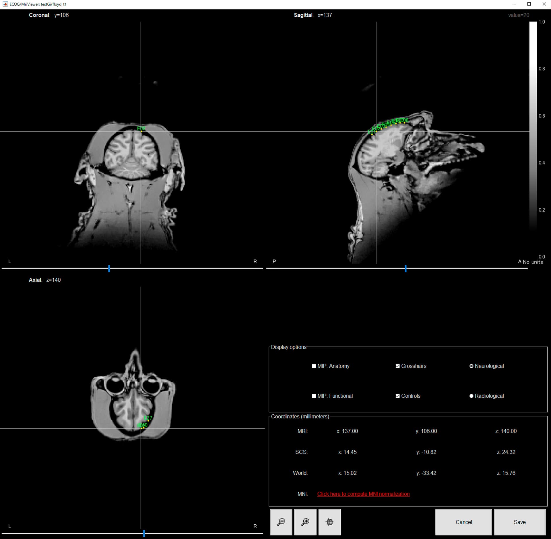
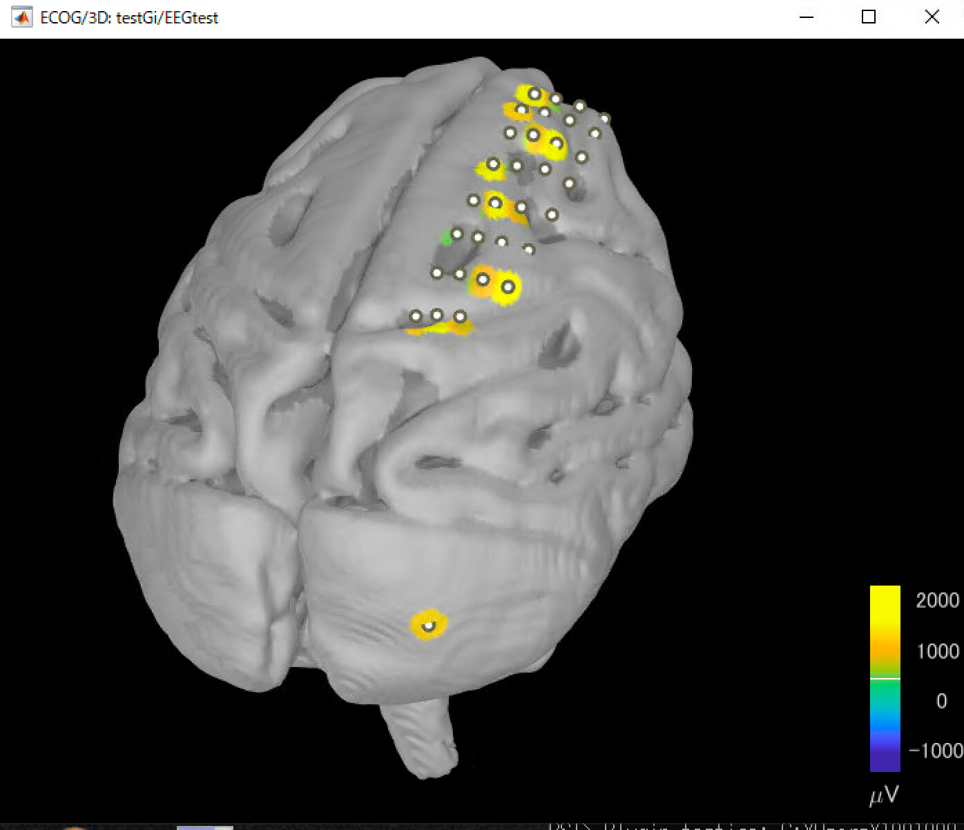
However, the area where the signal is displayed is too large for the electrode size.
As a test, one channel is placed at a distance from the other. You can see how large it is.
Can the signal spread be adjusted?
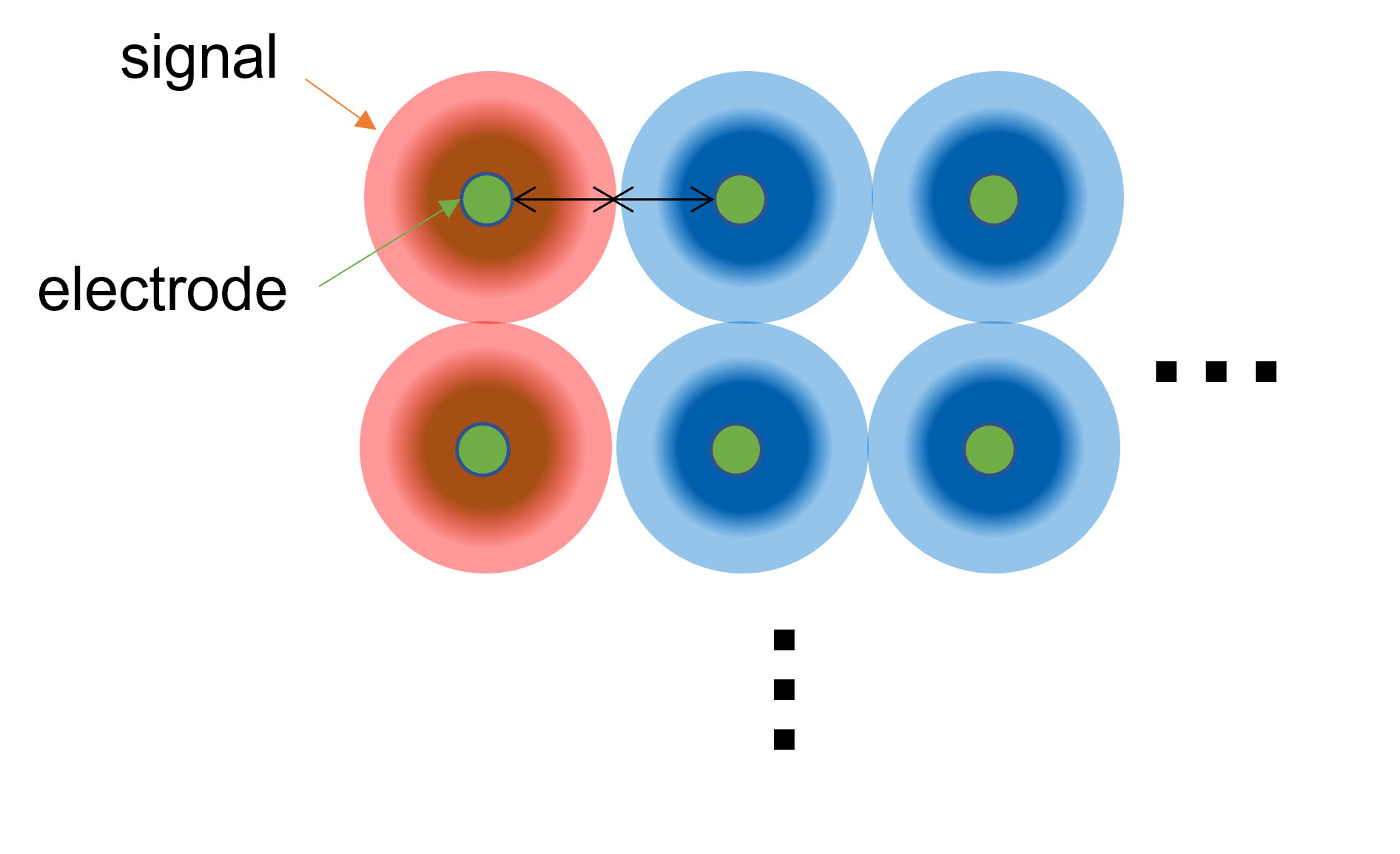
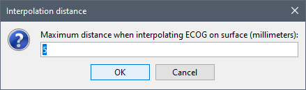
As @Francois says here, the parameter "excludeParam" probably determines the vertices to which the signal values are assigned, but is there any way to adjust this?
If < 0, exclude the vertices that are further from the absolute distance excludeParam (in millimeters)
Why should the colormap setting be changed?
The issue is the area where the signal is projected onto the cortex (the number of vertices to which the signal value is assigned), not the range of the signal value.
The parameter excludeParam controls for the maximum distance from the center of the contact.
The default values are set in panel_surface.m, it is 15mm for ECOG:
Your screen capture confirms that your channels are not correctly configured as ECOG.
If you used "ECoG" or did not change it from the default "EEG", make sure you set all the channels to "ECOG" (all capitalized).
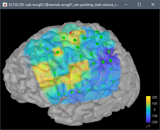
Rendering should look like this:
If you haven't read them yet, I recommend you follow the EEG, SEEG and ECOG tutorials before processing your own data:
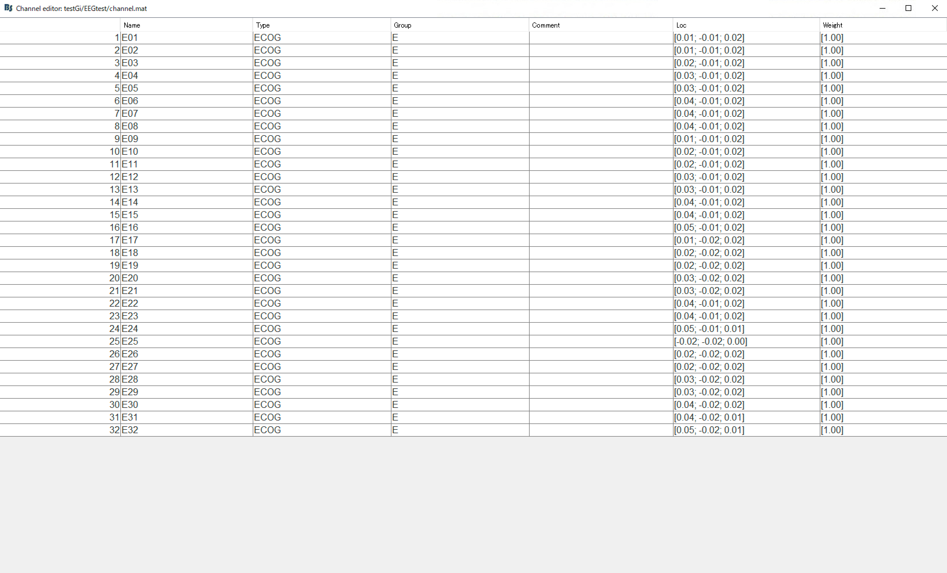
I think I correctly set the channels as ECoG.
Here are the steps I took for the ECoG data.
First, I select "import MEG/EEG" and then import a mat file because my ECoG data is defined as a MATLAB matrix (time * channel).
Second, I set the channel type as "ECOG" according to this.
Finally, I placed the electrodes on the MRI viewer and projected them onto the surface.
However, I have a request.
I am using the standalone version because do not have MATLAB.
Therefore, could you please compile the latest version and commit it?