Hello
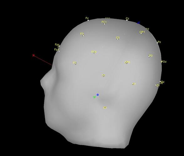
I'm analysing some EEG data using the default anatomy, and electrode positions from the ICBM152 ASA10-20 setup. (I used a 32 channel ASA waveguard cap for my recordings.) The location of the channels looks fine when I look at the arrangement with 3D views.
However when I load some EEG data and select the 2D sensor cap view, the 2D image seems like it is distorted i.e. not the full shape, even though it shows all the channels.
I'm concerned that this may interfere with SSP or ICA analysis, or later on source localisation? Could you clarify for me why this image looks distorted and whether it's a problem?
Many thanks
Luli


