I want to import a freesurfer volume atlas ( thalamus nucleis atlas, see below) in subject space for automatic SEEG electrode localization. Would you please help me about this? How can i import this volume as atlas in BST?
After importing the FreeSurfer segmentation folder as the subject anatomy, right-click on the subject > Import MRI.
Select the file format "Volume atlas (subject space)" if this volume was computed by FreeSurfer for this particular subject.
Select the file format "Volume atlas (MNI space)" if this a parcellation in MNI space (very unlikely from FreeSurfer).
I think you should answer Yes to the first question (Use the default FreeSurfer transformation). If the parcellation is not aligned correctly on the subject T1, then try other options.
Answer NO to the questions about registration and reslicing.
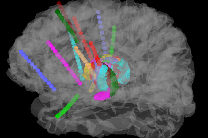
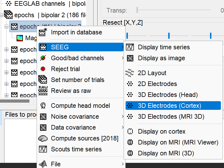
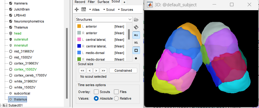
Thank you! I imported the atlas and created the surface successfully. And I want to show the SEEG contacts with functional data (amplitude of sEEG or so) in the new thalamus surface, but I find the new surface cannot be shown during displaying the SEEG contacts or sEEG data. So I wonder what was the problem?
However, if your goal is to display the ROIs defined in it as surfaces in 3D views, import it using the popup menu Import surfaces instead, with the file type "Volume mask or atlas".
I added a new option to add any surface to any 3D figure.
When you have an open 3D figure with the SEEG electrodes displayed in it, go to the Surface tab, click on the button "Add surface" on the top-right corner of this tab, then "Other", and finally select the surface to want to add to the display.
Hi Francois,
Thank you very much ! I have added the imported surface successfully in 3D views.
However, when I do the similar thing for the SEEG recordings, the additional suface cannot be shown, that is, nothing was shown after I added a new surface. So I wonder what's the wrong?
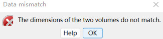
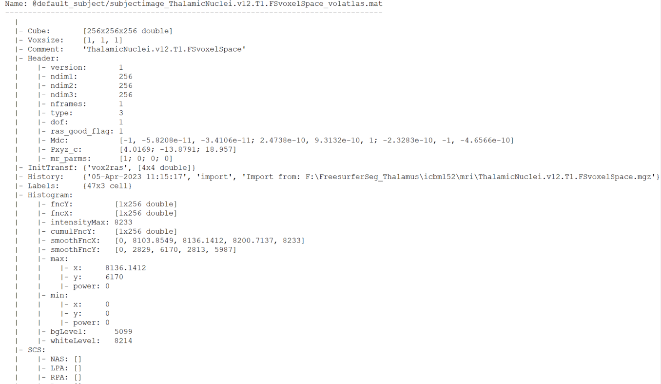
In addition, I want to use the thalamus atlas in the default anatomy, so I export the defalt ICBM mri image and used it for Freesurfer rencon-all parcellation and got the atlas. But when I imported the new thalamic volume atlas (subject space), the atlas cannot be overlayed on the default MRI. The error was as below.
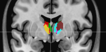
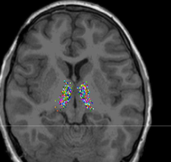
when I imported the volume atlas (MNI space), the atlas could be overlayed but it seems the atlas was not well aligned to the MRI.
But when I imported the new thalamic volume atlas (subject space), the atlas cannot be overlayed on the default MRI. The error was as below.
The logic of exporting the ICBM152 T1 MRI and processing it with FreeSurfer should work.
However, FreeSurfer rewrites the MRI in a 256x256x256 volume (T1.MGZ) and everything is aligned on this, not on the original volume exported from Brainstorm. So you can't import the thalamus parcellation directly in your default anatomy folder.
If you want to do this, you have to create a new subject with the option "Default anatomy: NO", import the full FreeSurfer output folder as the new subject anatomy, and finally import the thalamus atlas.
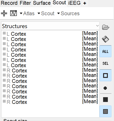
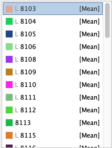
Hi, Francois I am also working on thalamic segmentation. I would like to ask the way to add this above mentioned free surfer based ThalamicNuclei.v12.T1.FSvoxelSpace .mgz as an atlas to the MRI. I tried the suggested way and successfully could make 3D picture with a surface of thalamus and electrodes. However when I try to project to the MRI as an atlas (by importing as a Volume), it seems that the "Label" was blank. Therefore I uploaded those according to the freesurufer LUT, to the Label, but seems the interpolation is not working. Could you please suggest me how to solve this?
Thank you very much for your help!
Hi @anaskhan07, since this segmentation of thalamic nuclei are not part of the standard output of recon-all it labels are not properly handled in Brainstorm. Note that the label names are not saved in the volume atlas.
However, as mentioned in the post above, the labels can be gotten from the FreeSurferColorLUT.txt file, as such this is the direct link to these labels: