I'm working on the neural representation of facial expressions (like fear), so I need to localizing subcortical sources, especially amygdala. I tried to follow the DBA tutorial and faced some problems.
In this tutorial, the default orientation constraint of cortex is constrained, and the default orientation constraint of amygdala is unconstrained.
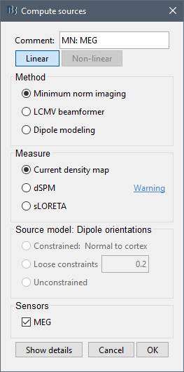
When I compute sources with mixed model, the tutorial says
Select all the subjects (except the empty room recordings), right-click > Compute sources.
Select Minimum norm (wMNE) and leave the other options to their default values.
I am wondering that whether this source is constrained or unconstrained, or mixed.
How can I project this source on default anatomy?
Based on the tutorial,
Right-click on "aseg atlas" > Less vertices > 15000 vertices.
Double-click on "aseg atlas_15000V"
Select the amygdala, the thalamus and the hippocampus, and create a subatlas.
Merge the cortex with the selected structures. Rename the new surface in "cortex_mixed".
Open the cortex_mixed surface and create a new atlas "Source model options".
Set the set modeling options to "Deep brain" for all the structures.
I operated these steps on Default anatomy and every subject.
I estimated sources for every run and made a weighted average for each subject. Then I processed baseline normalization (Z-score transformation), Rectify(absolute values) and Project(project on default anatomy: surface).
Are these operations correct?
I am wondering that whether this source is constrained or unconstrained, or mixed.
It depends on the forward model you are using. If the comment of your head model says "mixed", then when estimating the sources you can compute only a "mixed" head model, the options about source orientation are disabled in the "Compute sources" options. Depending on the DBA atlas, some regions will use constrained dipoles locations and orientations and others unconstrained dipole orientations. See the reference articles for more details.
I estimated sources for every run and made a weighted average for each subject. Then I processed baseline normalization (Z-score transformation), Rectify(absolute values) and Project(project on default anatomy: surface).
Projecting mixed models to a template works but is not accurate. The cortex registration between subjects is reliable (based on the registered spheres of FreeSurfer: https://neuroimage.usc.edu/brainstorm/Tutorials/CoregisterSubjects), but the subcortical structures are not. The various volumes are not correctly co-registered - you can see the results of this registration when running the projection, and therefore the interpolation of source maps between subjects is not very precise.
If you want to go on with the your pipeline with the mixed head models: if you are getting errors when projecting the source maps, update Brainstorm. I've just found a small bug that is now fixed.
Thank you for the reply! I’ll try volume source models.
After the operations I have mentioned above, I exported the sources to SPM (Export to SPM8 (volume)) to do group analysis. Since the subcortical data is not accurately registered, shall I separate the cortical data and the subcortical data, and only analyze the cortical part?
Can [Export to SPM12 (surface)] only export the cortical part?
I don’t see how you could split the various elements of your source results after computing them.
It is simpler to recompute new results with a volume head model or with only cortical structures.