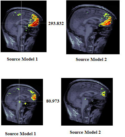
Although I see the similar activation in both the models but the intensity is different. Please see the figure below. Why is this happening?
This is difficult for us to know what you did differently the two times you computed these maps.
Note that the "intensity of the color" does not mean anything without knowing what the bounds of the colormap are. Change the maximum displayed value in a figure, it changes the colors.
https://neuroimage.usc.edu/brainstorm/Tutorials/Colormaps#Color_mapping
Are these some power measures, computed for a specific frequency band?
I extracted the scouts and overlayed two scouts, these look very noisy. I followed the preprocessing tutorial and removed eye-blinks, ECG artefacts using SSP (as you can see in the below figure)
Ongoing/resting recordings typically can't be explored by looking at them time point by time point like you do here. You need to compute some measure from these recordings (power in a frequency band, connectivity between two sensors or ROIs, etc), and then compare these timeless measures across conditions or groups of subjects. See this tutorial:
https://neuroimage.usc.edu/brainstorm/Tutorials/RestingOmega
Going through the SSP tutorial, it says that SSP projectors will be applied on the fly to the data, but I can still see the blink artefacts. I followed the steps in the tutorial. Is there anything I am missing?
The SSP projectors you computed are probably not adapted or sufficient for removing the blinks correctly. Try refining the list of blinks you have given the SSP computation, changing the selection of components that are removed, or adjust the parameters of the SSP computation manually.
https://neuroimage.usc.edu/brainstorm/Tutorials/ArtifactsSsp#Troubleshooting
https://neuroimage.usc.edu/brainstorm/Tutorials/SSPCookbook
You can also try with ICA:
https://neuroimage.usc.edu/brainstorm/Tutorials/Epilepsy#Artifact_cleaning_with_ICA
I used PSD+Scalp topography to identify the eye blink artefact and noticed activation between 2-7Hz indicating the EOG. Will SSP remove this frequency component from the recordings?
Indeed, the eye movements will be most visible between 1 and 7 Hz:
https://neuroimage.usc.edu/brainstorm/Tutorials/BadSegments#Automatic_detection
The objective with SSP/ICA approaches is to remove specific components (topography representative of the artifact) without having to remove all the frequency band in which the artifacts are visible.
If you have no interest in what is happening below 7Hz in your recordings, you could apply a low-pass filter at 7Hz at the very beginning of your analysis.
It could help you to start by some additional background readings on EEG/MEG signal processing before trying all these procedures directly on your recordings:
https://neuroimage.usc.edu/brainstorm/Tutorials#Background_readings