Weird results of PSD when project to default antomy

,

Dear Brainstorm forum,

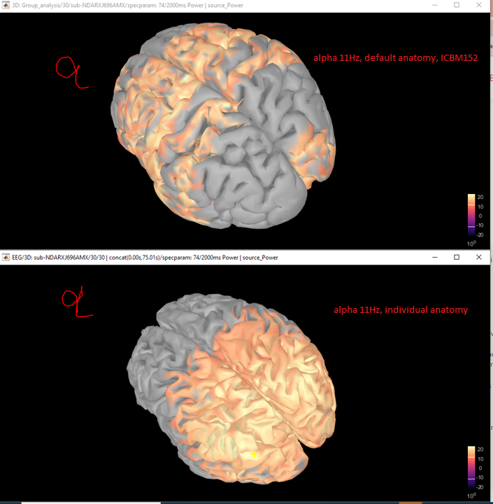
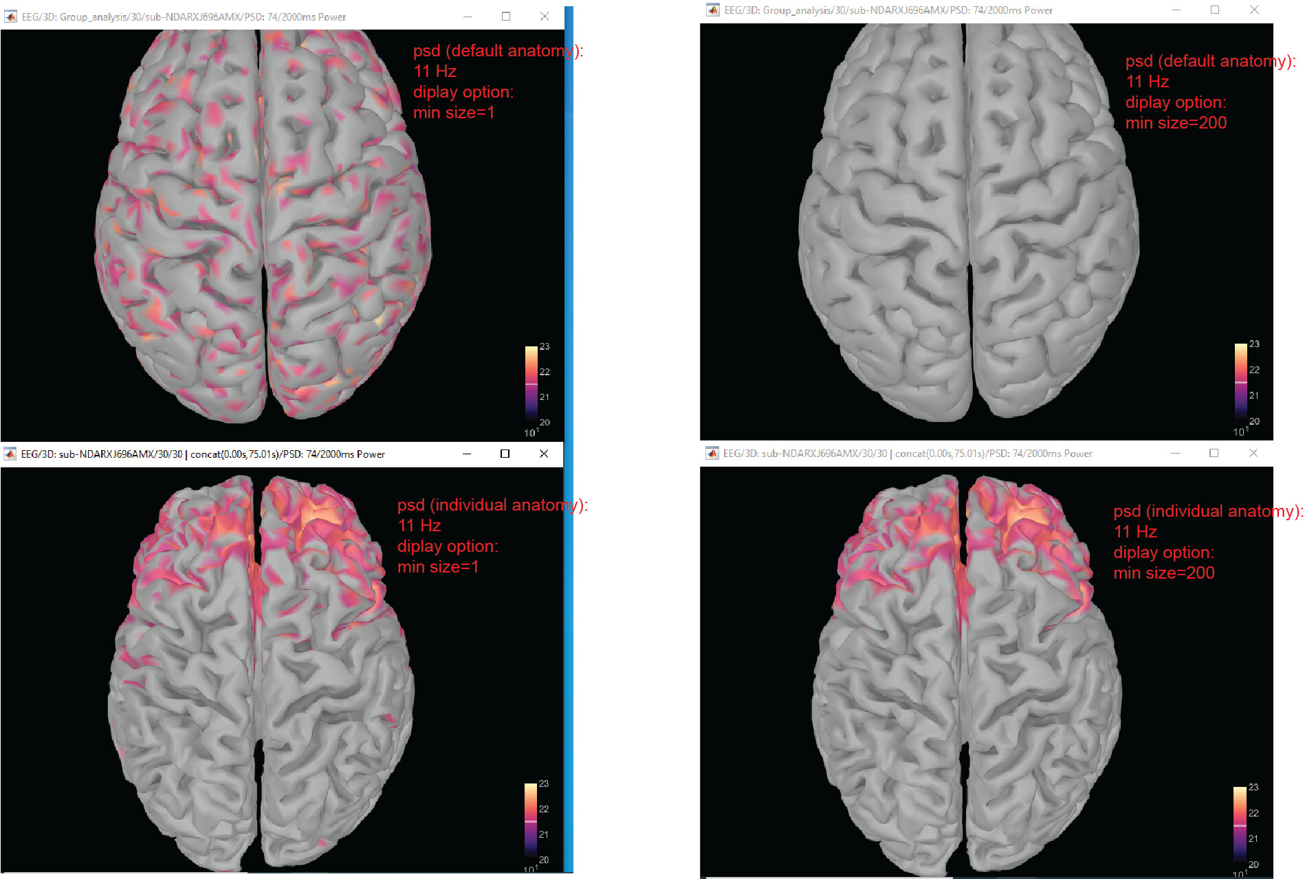
I am working on PSD EEG-reconstruction using individual anatomy. After computing PSD (+fooof), I project the PSD result to default cortex (ICBM152 cortex_15002v), however, I get a very confused map (alpha band at resting EEG condition, attached figures). Basically, alpha peaks present in ocipital area in individual anatomy but it expand to everywhere in default cortex.

I don't know which steps I did wrong. Which steps I need to double check?
Thanks.
Best,
Xiaoyue

  1. Select each figure (clicking on the black part) and check that both figures are using the same Measure and spectparam options in the Display tab (main Brainstorm window).

  2. If both images have the same visualization parameters. Try the project to the subject's PSD (before fooof) to the default anatomy, and check if projection is correct.

Hello Raymundo,
Thanks. However, the psd projection is also strange, see the screen-cut. what else can i do?
By the way, the subject is 21.19-year-old. EEG recording cap is EGI-129.
Thanks again.

Xiaoyue