Digimouse: Data Acquisition
Overview | Data Acquisition | Registration | Creation of Atlas | Simulations | Download | License
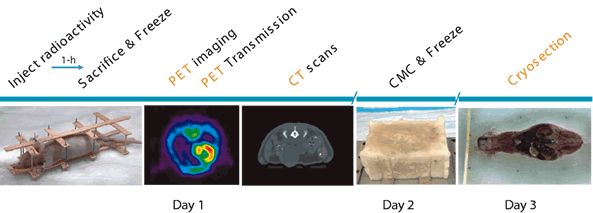
We created high quality images of PET, CT and cryosection slices markers readily apparent for later image registration that would not compromise the image acquisitions. We accomplished this by creating a wooden framework to which both the mouse and markers were attached. The markers consisted of small bore teflon tubing filled with black ink and F-18. The mouse was frozen by liquid nitrogen immersion after being sutured to the framework and was kept frozen during imaging of PET and CT by blowing cold nitrogen gas over the mouse. Following PET and CT imaging, the mouse and the framework was packed in CMC and froze it overnight at -20C. The next morning the cryostat was set up to cut the sections thoughout the entire mouse.
- PET images were acquired in one bed position for 15 minutes in a Concorde P4 microPET following 60 minutes uptake. A combination of F-18 ion (for bone uptake) and FDG (for heart, soft tissue and bladder uptake) was used and images were reconstructed using filtered backprojection to 2.2 mm resolution.
- CT images were acquired in 2 bed positions using Imtek microCAT system (Imtek, Knoxville, TN) and reconstructed into 512 cubed matrix for a pixel size of ~100um.
- Cryosections were cut at a thickness of 50 um and imaged using a Nikon Coolpix 5700 digital camera, with an in-plane voxel size of ~38.8um.
Current configuration does not allow embedding of the file sequence_new.gif because of its mimetype image/gif.: sequence_new.gif
The Raw Data of coronal PET, CT and Cryosection slices are:
|
|
|
|
|
PET |
CT |
Cryosection |


