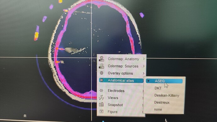
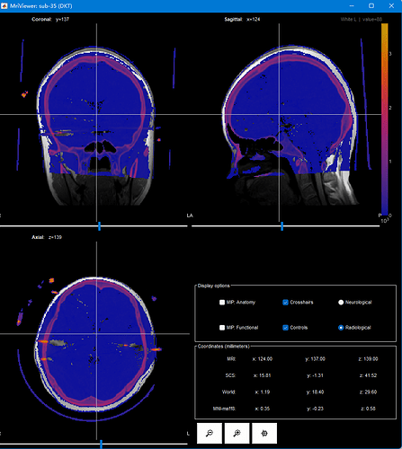
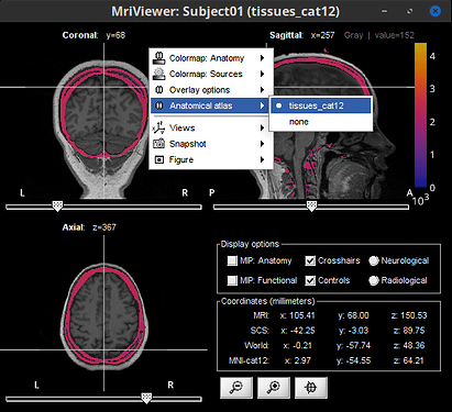
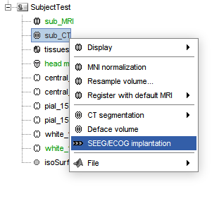
I am attempting to locate the SEEG electrodes using the updated Brainstorm software (version from 25-Oct-2024), but I have encountered difficulties. The task cannot be performed using the raw CT scan or the registered CT scan without reslicing. However, when I use the resliced CT scan, the atlas cannot be overlayed. Can you please provide guidance on how to resolve this issue? Thank you for your help!
This is the error info when I using the only registered CT to perform electrode location:
***************************************************************************
** Error: The dimensions of the two volumes do not match.
***************************************************************************

error using getappdata
value must be handel
error panel_ieeg (line 31)
eval(macro_method);
error
tree_callbacks>@(h,ev)panel_ieeg('DisplayChannelsMri',filenameRelative,DisplayModReg{iMod},iVolAnat(iAnat),1) (line 981)
gui_component('MenuItem', jMenuAlign, [], [strType 'Edit... (MRI Viewer: ' sSubject.Anatomy(iVolAnat(iAnat)).Comment ')'], IconLoader.ICON_ALIGN_CHANNELS, [], @(h,ev)panel_ieeg('DisplayChannelsMri', filenameRelative, DisplayModReg{iMod}, iVolAnat(iAnat), 1));I find the new version 241030, I will test this.
The error still exist in version 241030.
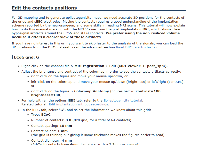
Did you use the following tutorial for coregistration? https://neuroimage.usc.edu/brainstorm/Tutorials/Epileptogenicity
This is correct, to plot the MRI and CT in the same figure, they need to have the same dimensions (this is to say the CT was resliced to match the MRI volume).
You can verify the volume size for each of the 3 files (MRI, CT and Anatomical atlas), with right-click on the file in the Brainstorm database, then File > View file contents and check the dimensions of the Cube field.

- Can you provide more information about the imported MRI and its CT?
- Can you display the volume atlas only on the MRI?
Please note that the volume atlas will not be shown in the plots with MRI and CT, as the CT is already an overlay layer. However, you can still get the voxel labels. As shown in the figure below. MRI (gray scale), CT (color scale), Anatomical atlas (not visible), Label current voxel (top right legend)
Sorry for the late reply. Thank you so much!
I understand the differet volume dimention between T1 and CT. And I can display the volume atlas on MRI. But, in previous versions, I was able to use the registered CT alone for electrode localization, without the necessity of overlaying it on the T1 image.
The CT must be also coregistered, and resliced, so it is in the same space than the MRI.
Check this link:
https://neuroimage.usc.edu/brainstorm/Tutorials/IeegContactLocalization#Import_the_anatomy
I appreciate your perspective. However, according to the tortuial, it is recommended to use the non-resliced CT for electrode localization. Unfortunately, in the version dated 30-Oct-2024, I am unable to utilize the non-resliced CT to adjust the electrode's location.
https://neuroimage.usc.edu/brainstorm/Tutorials/ECoG
Hi @longzhou, thank you for the detailed information, now I fully understand and can replicate the issue you are facing.
Indeed, this was introduced in a recent update on the iEEG localization methods
For the moment, a walk around is to import the CT volume as MRI (with the Import MRI) option. And make this CT volume the only MRI (deleting the T1). Then you should be able to continue with tutorial as described. After the iEEG localization you can import back your T1 MRI.
@longzhou, apologies for temporary disruption to your workflow, as you know we are always adding new features to improve Brainstorm capabilities. However, these improvements often come with new bugs (like in this case) and then bug fixes. This mechanism works thanks to user like your yourself that report these changes.
![]() @chinmay.chinara, this is high priority please add the feature of allowing iEEG localization in one volume only (MRI or CT) to the list of things to improve in iEEG dev
@chinmay.chinara, this is high priority please add the feature of allowing iEEG localization in one volume only (MRI or CT) to the list of things to improve in iEEG dev
Great Toolbox!!!!
@longzhou, we have improved the ways to start the SEEG/ECOG implantation.
Now, it is possible to start it directly from your CT volume (with right-click on it). With this option only this CT volume will be used (thus avoiding the trouble of having a MRI volume of different size)
Please update your Brainstorm instance to get this feature.
