
Cortical Delineation Protocol
IntroductionCortical surface-based registration methods differ mainly on the features, or similarity metrics, they use when aligning cortical surfaces. One general approach uses manually or automatically defined landmark contours to constrain the registration. In this page we describe a set of landmarks that can be used for cortical registration. The protocol consists of 26 sulcal curves that consistently show in normal brains, and are distributed throughout the cortical surface. Tracing is performed using the software BrainSuite. ContributorsThe cortical delineation protocol was developed by Hanna Damasio at the Dornsife Cognitive Neuroscience Imaging Center and Brain and Creativity Institute at the University of Southern California, in collaboration with Dimitrios Pantazis, Richard M. Leahy and David Shattuck. For more details see: D. Pantazis, A. Joshi, J. Jintao, D. Shattuck, L. Bernstein, H. Damasio, R.M. Leahy "Comparison of landmark-based and automatic methods for the registration of the cortical surface", NeuroImage, in revision
Demonstration of cortex delineation using BrainSuite (video file) Curve Delineation ProtocolA thorough description of the protocol curves, with tracing details, figures and relevant information, is given in the pdf file:
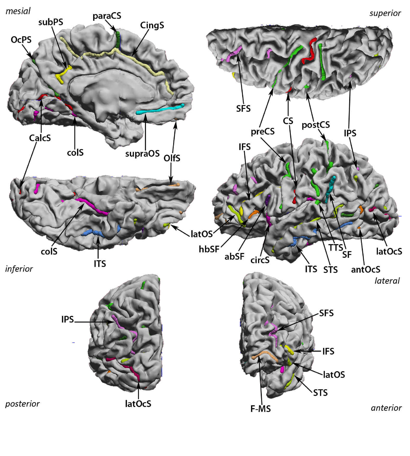
1) central sulcus (CS) RequirementsTo perform cortical registration using our curve protocol, you need: 1) Download BrainSuite a magnetic resonance (MR) image analysis tool designed for identifying tissue types and surfaces in MR images of the human head. 2) Download the curve protocol file: sulcal_protocol CitationsTo cite this protocol: D. Pantazis, A. Joshi, J. Jintao, D. Shattuck, L. Bernstein, H. Damasio, R.M. Leahy "Comparison of landmark-based and automatic methods for the registration of the cortical surface", NeuroImage, in revision To cite BrainSuite: Shattuck DW and Leahy RM (2002) "BrainSuite: An Automated Cortical Surface Identification Tool", [invited article] Medical Image Analysis, 8(2):129-142 Shattuck DW, Joshi AA, Pantazis D, Kan E, Dutton RA, Sowell ER, Thompson PM, Toga AW, Leahy RM (2009) Semi-automated Method for Delineation of Landmarks on Models of the Cerebral Cortex, Journal of Neuroscience Methods 178(2):385-392. BackgroundFor this protocol of BrainSuite all sulci are traced on the brain's midcortical surface. On occasion, when sulci are very deep, they may be shown on a gray/white junction surface (but the curves will have been traced on the midcortical surface). All sulci are traced at a 0.5 of "stickiness". When jumping over gyri which interrupt the course of a sulcus it is best to remove the "stickiness' because it facilitates the placement of the curve. It is also advisable to turn the surface view in such a way that the dropping of a point is done perpendicular to the deep surface where the point is meant to be dropped. The different sulci curves cannot touch one another, there has to be a gap between them. This is important to keep in mind when, on occasion, some sulci actually cross other sulci. For alignment purposes they have to be kept separate. The sequence in which the sulci are traced is arbitrary. Here, and in the actual protocol in BrainSuite, the sulci are ordered by lobes, starting with the Frontal, continuing with the Temporal, the Parietal and finally the Occipital. In the Frontal and Parietal lobes I start with the dorsolateral views, move to the mesial views, and to the inferior view (in the frontal lobe). The Temporal lobe starts on the dorsolateral view and gradually proceeds to the mesial view. The occipital lobe starts with the mesial view because this is the view where its major sulci are seen, and finishes with the dorsolateral view. For a general overview, the first images show the brain in straight lateral, mesial and inferior views, with all sulci traced and identified with the abbreviations of the sulci names (all introduced in the text for the individual sulci). The image using the pial surface also has the major gyri labeled. Recommended sources of further information for sulci (and gyri) identification: H.Damasio, Human Brain Anatomy in Computerized Images , 2 nd Edition, Oxford University Press, 2004; H.M.Duvernoy, The Human Brain: Surface,Blood-Supply, and Three-Dimensional Section Anatomy , 2 nd Edition, Springer Verlag, 1999; M.Ono et al., Atlas of the Cerebral Sulci , Thieme Verlag, 1989. |
|
USC Biomedical Imaging Research Lab © 2009, designed by Dimitrios Pantazis |