I am following this tutorial: https://neuroimage.usc.edu/brainstorm/ExportSpm8
And I encounter an error: after exporting the MRI and opening SPM, I can't find the exported .nii file in the folder that it is saved on the SPM batch editor.
Hi,
I solved the problem, I was on the Directory browse, not the file browse.
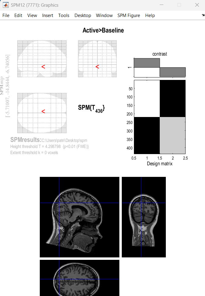
However, once I finished that and get to the end of the tutorial, I find that I get a blank analysis:
The process works fine, so the issue seems to be with what you are exporting
Please provide context on the files that you are exporting, as well as the parameters for the process. Feel free to add the screenshots with how the data looks on Brainstorm, to verify there are sources to export.
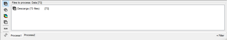
I believe that it has something to do with the process on Brainstorm, because on the tutorial the files to process remain the number in between brackets, in my case 73, but on the tutorial 101, I upload both screenshots below:
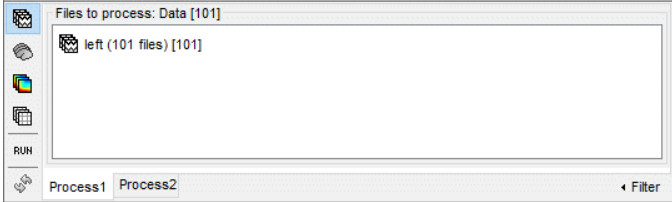
In the tutorial, it is also mentioned to select "Process sources", but if I do that, the number of files triplicate while on the tutorial it remains the same 101 number until the end of it, even on the spm process:
This is correct as you cannot export sensor time series as MRI volumes.
When you click on Process sources, the files to be processed as the sources belonging to those sensor time series. If they are triplicated, this suggest that you have 3 source estimates per sensor time series.
The process is working fine on our side and for other users. So, it may be a case that is not well utilized on your side. Please check carefully the data that you have, and what you expect, if there are questions about that do not hesitate in sharing them here.
Do not assume, you have all the elements to verify it
However, I tried several times to run the spm process and I still get a blank result.
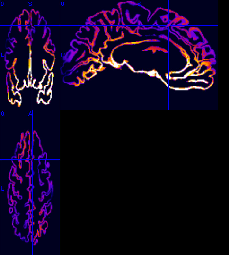
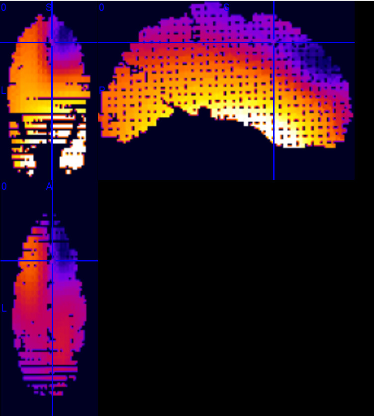
With the MRIcron I can also see that the files are "divided" as I show below and get both the surface and the filling of the activation. Is this normal after exporting the Brainstorm process?
Sources estimated in the cortical surface space. I.e., dipoles are only distributed on the cortex. Thus, when exported only voxels on the cortex have values.
Sources estimated in the entire brain volume space. I.e., dipoles are distributed on a 3D grid in the entire brain volume. Thus this source grid is interpolated to the MRI volume, since the source grid is less dense than the voxel "grid", then you see blank spaces.
There are not "correct", surface or volume source grids can be used to estimate brain activity. Which one you use depends on your research question.
Please check the tutorials for (surface) source estimation, and the volume source estimation. There are important references at the end that you can consult to make these concepts clearer.
Good day, my research purpose is to compare the epilepsy source on both intracraneal and surface analysis performed on Brainstorm.
However I am stuck because SPM does not recognize the vowels, and I get this error: