I'm working on source localization from participants with individual MRIs and EEGs.
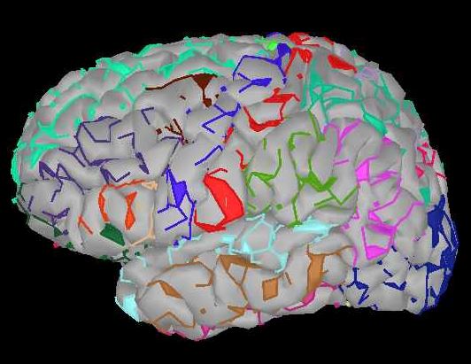
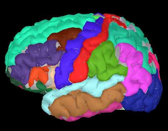
In looking at scout options, I'm visualizing the templated atlas's onto cortex. The templated ones look great (e.g., DKT_1, attached here). However, when I try to create from the subject's anatomy, the image looks less complete (DKT_2). Smoothing the image looks better, but I'm worried that I am doing something wrong. ANy thoughts? THank you!
If the MRI segmentation method that was used did not return the cortical parcellations that you want, a way to get them would be to project them from the Default anatomy to the Subject anatomy.
Display the cortical surface from the Default anatomy
Make a copy of the Atlas you want to project with the option Atlas > New atlas > Copy current atlas
Select the copy of the desired Atlas and select all the Scouts
Use the menu Scout > Project to... > SubjectName > SurfaceName
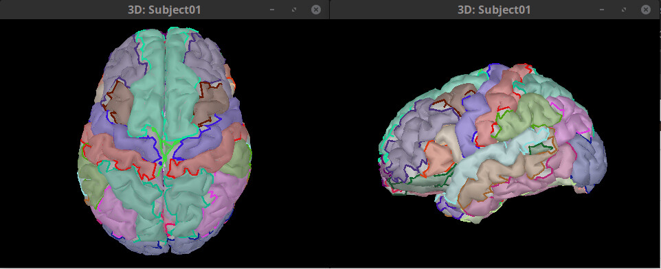
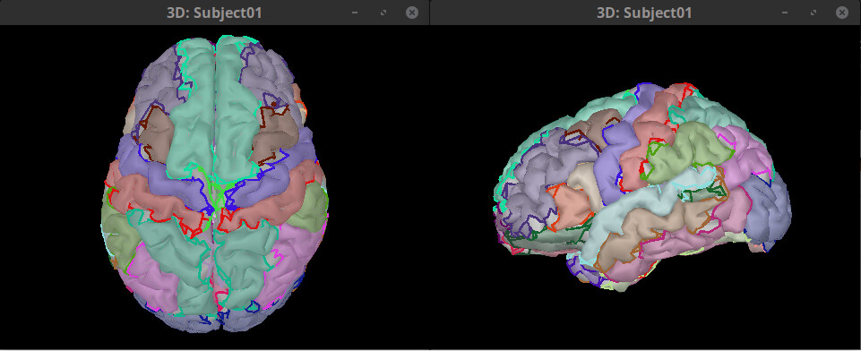
Example of results:
DK atlas imported from MRI segmented with FreeSurfer
Thank you! That makes sense, although I'm confused by the alternative.
I'm trying to use brainnectome_leadDBS to identify scouts, but I can't see it within the 'scouts' tab unless I use the 'from subject anatomy' option.
Is there a way to get the non-templated (e.g., schaefer, brainnectome) to be options within the scout tab alongside the templated (e.g., DKT, brodmann, etc)?
I had previously added the brainnectome_leadDBS parcellation in the anatomy tab.
In Brainstorm, the term Atlas is often used for two different things:
An anatomical atlas. These are volumes where each voxel has a label. This type of atlas are shown in the Anatomy view with this icon:
A collection of scouts. First, a scout is a set of vertices that were used to estimate sources. There are surface and volume scouts, since sources can be computed in surface or volume grids. Thus there are surface and volume Atlases. More details in the head model tutorial (below). Scout and These are save inside surface files. Even if source space is a volume grid, there is not a one-to-one equivalence between a voxel (anatomy volume) and a vertex (volume grid). https://neuroimage.usc.edu/brainstorm/Tutorials/HeadModel
This is an anatomical atlas. So, when you try to import it as an surface Atlas (made of surface scouts), the surface vertex are converted to MNI coordinates, and the label is search in the anatomical atlas. As you saw this does not always give good results. You could try to do it directly in the Scout tab as Atlas > Load atlas... and select the anatomical atlas .nii file as Volume mask or atlas.
Ohhh! Okay, this was incredibly helpful. I see my error. Thanks Raymundo.
Follow-up re brainnetome atlas. I see in this post a few years ago (below), if we download and use FSAverage_2020 as a template, I now see multiple other scouts available (including brainnectome). these appear to be cortical and not volumetric. is this an appropriate approach?
Now I'm realizing that this approach just overrides the individual's MRI with the FSAverage MRI. I believe I need to map the brainnetome atlas onto the individual's MRI in free surfer, prior to brainstorm, and then when i upload anatomy to brainstorm, the brainnetome atlas should be available. right?