I have already done the source analysis with Brainstorm and now I want to download the result with the epileptic source identified so I can enter it to an external Matlab app and perform a cat12 analysis, but I encounter several problems:
I don't know which item of the Brainstorm dropdown I have to download, because I know that for the cat12 analysis, the item must be in .nii format.
I tried to download the Dipoles EEG item, which is an MRI, but the cat12 analysis returns an error. I am not sure that this is the one I am looking for.
Once you have computed the volume source estimation (either MN or dipole modeling), you should have a file with the (sources) results: or
Just drag-and-drop such file to the Process tab and select the process File > Export to SPM8/12 (volume), use the desired preferences, and click on Run this will create the NIfTI file.
Is this what it is expected to be?
I am not sure that i will be able to perform any analysis on the data extracted from here.
The intention is to extract the source information obtained after the sLORETA analysis to compare it between several files and be able to identify the area that is shown in both.
Any ideas?
Well, that depends on your data and the analysis, it is not possible for us to say without further context.
It seems you are exporting a sources estimated on the (cortex) surface, so, the exported NIfTI volume is only the cortex. You may want to estimate sources in the volume space, the volume source grid will be interpolated to the MRI volume.
And how can I estimate sources in the volume space?
I expect to get an MRI with the epileptic source highlighted, as in Brainstorm, but saved as a regular MRI that can be analyzed by cat12 in an external Matlab app.
If you are seen the sources in the MRI space, it is likely you estimated the sources in a volume grid. Please share some screenshots of what you see in Brainstorm, so we can help you out better.
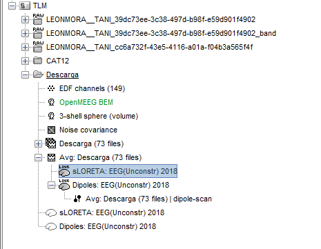
As you can see in the shared screenshot of the database, there are two head models in your database:
OpenMEEG BEM
3-shell sphere (volume)
Since the OpenMEEG BEM head model does not have the indication "(volume)", it suggest that the selected source space was the cortical surface.
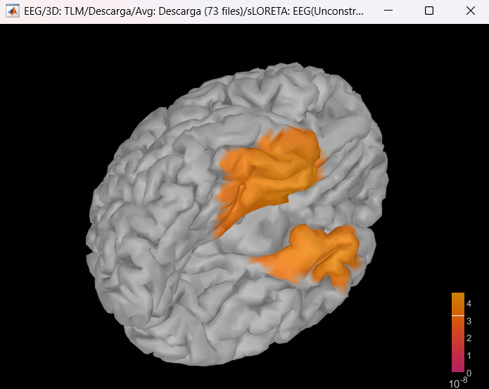
Then, the sLORETA estimation seems to be computed with the OpenMEEG BEM thus computed for the surface space, this is confirmed with the plot you share where sources are only distributed on the cortex.
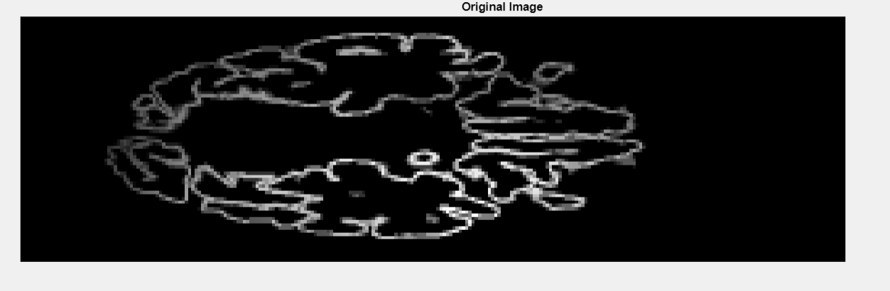
Finally, if you export these cortical sources to a NIfTI file, only voxels that correspond to the cortex will have value, which is what you observed in the first figure of this thread
Please check these tutorials on head model and (surface space) source estimation to understand better the source spaces:
Thank you for your response.
I computed sources after selecting the 3 shell sphere volume and selected sLORETA.
What I get after that is a 3 view MRI with the source stimation.
My concern is that i may have some trouble when performing the cat12 analysis becouse of the 3 views.
Not really. What you get is the source density for each vertex in a 3D grid. This 3D grid is then interpolated to the MRI voxels. The 3-view MRI that you see, is just the sources on these slices, the data is defined for the entire volume. Thus it should be possible to analyze it with CAT12 as a volume.
Please read the section Using unconstrained sources in the first reply: