
Central Sulcus
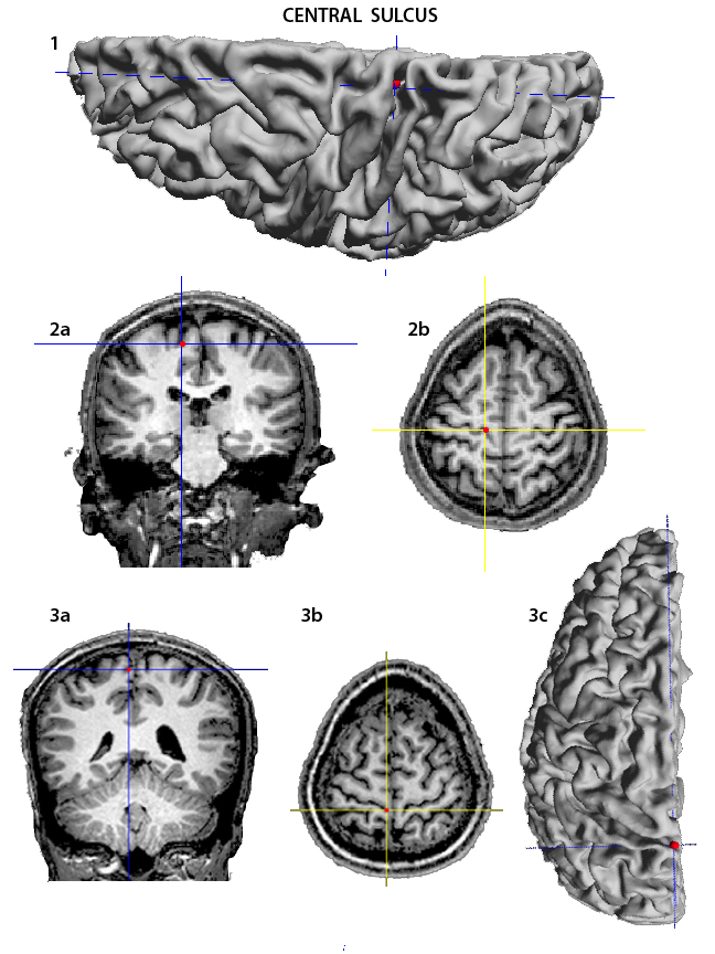
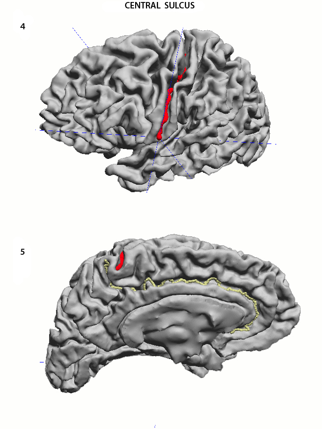
The Central Sulcus (CS) separates the frontal lobe from the parietal, and constitutes the posterior limit of the precentral gyrus. It can be found on the lateral surface of the hemisphere, where there are 3 parallel sulci running superior to inferior. The CS is the middle one. It is most often a continuous sulcus that starts at the interhemispheric fissure and runs inferiorly and anteriorly towards the Sylvian Fissure. It may or may not reach it. The superior end may actually be on the mesial surface of the hemisphere. For the purpose of alignment of different brains using the sulci as constraints, the superior starting point should be considered on the dorsolateral surface, close to the midline interhemispheric fissure. Begin by aligning the hemisphere perpendicular to the screen, looking at the edge of the interhemispheric fissure (1), and drop the first point close to the midline. Check in the coronal and the horizontal slices that the point is in fact on the dorsolateral surface (2a - b). In 3a-c are the images that show an initial point dropped on the mesial surface of the hemisphere. This is correct anatomically. However, because of the reasons given above to align different brains to each other using the sulci as constraints, it is preferable to restrict the sulcus to the dorsolateral surface. Move the hemisphere so as to see the full dorsolateral surface, and drop the endpoint of the CS (4). The mesial view of the hemisphere can help in the identification of the CS. Once the Cingulate Sulcus is identified together with its superior and posterior end (the ascending ramus of the Cingulate Sulcus), the CS is the small sulcus anterior to it with an antero-posterior direction (5).
|
|
USC Biomedical Imaging Research Lab © 2009, designed by Dimitrios Pantazis |