
Pre-central Sulcus
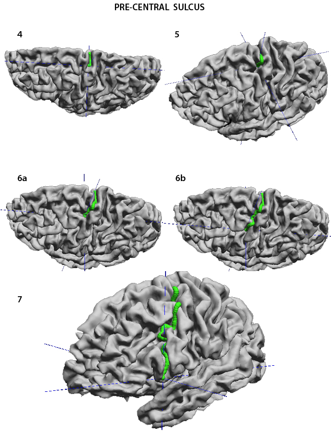
The pre-Central Sulcus (preCS) forms the anterior limit of the precentral gyrus. It is parallel to the CS and is immediately rostral to it. It also starts at the interhemispheric fissure and runs inferiorly and anteriorly towards the Sylvian Fissure. It is very often subdivided into two or more segments. Drop the first point of the curve high in the drsolateral surface of the hemisphere, close to the interhemispheric fissure (1), and check the position in the coronal and horizontal slices (2a-b), as was done for the CS. Then proceed to the end of that segment (3). To jump over the gyrus interrupting the sulcus select no stickiness which helps direct the trace exactly where it should go (5). Do the same to the next interruption (6a-b), and continue to the end of the sulcus (7). The surface image has to be rotated from drop to drop so as to maintain a view that is essentially perpendicular to the place where the next point is dropped. The beginning of the preCS can have two small branches in the form of a V . Either can be chosen, but it is important to maintain consistency across subjects. I chose the posterior sulcal extent (8), because it seems to be the more consistent course for this sulcus.
|
|
USC Biomedical Imaging Research Lab © 2009, designed by Dimitrios Pantazis |