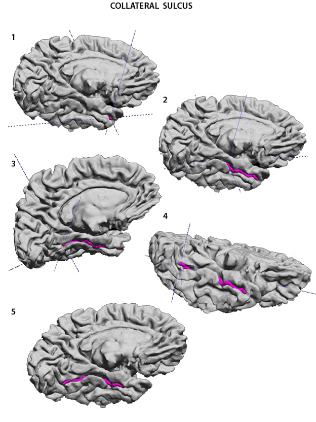

Collateral Sulcus
The Collateral Sulcus (ColS) is yet another anteroposteriorly running temporal sulcus. It is the most mesial temporal sulcus and provides the lateral limit of the parahippocampal gyrus. The ColS is best seen with the surface rendering tilted halfway between a mesial and an inferior position (1). The anterior end often merges with the rhinal sulcus to form the antero-medial border of the uncus. Here, to facilitate the marking of the curve, we include the rhinal sulcus a the most anterior segment of the collateral sulcus. Drop the starting point here (1). The ColS runs posteriorly paralll to the hippocampal fissure (3). It often splits into two rami which continue into the occipital lobe. The lower, or more lateral ramus (5) will separate the lingual and fusiform gyri in the mesial occipital lobe. The sulcus should be terminated at the posterior end of the temporal lobe, or continued into the occipital lobe chosing the lateral terminal branch. Once again, the importance is to be consistent across subjects.
|
|
USC Biomedical Imaging Research Lab © 2009, designed by Dimitrios Pantazis |