|
Size: 4070
Comment:
|
Size: 8624
Comment:
|
| Deletions are marked like this. | Additions are marked like this. |
| Line 4: | Line 4: |
| Brainstorm orients most of its database organization and processing stream for handling anatomical information together with the MEG/EEG recordings. The introduction tutorials start with the import of the T1 MRI of the subject, and this anatomy seems mandatory everywhere. These choices were made because the primary focus of Brainstorm was to estimate brain sources from MEG/EEG, which ideally requires an accurate spatial modelling of the head. If you don't have access to anatomical images of your subjects or if you are not interested in source reconstruction, Brainstorm will still require that you explicitly define an anatomy; in those case you would use an anatomy template. In the case of group analysis at the source level, you would also use a template on which you would project of the individual results. Several templates are available, with a large preference for using the MNI ICBM152 package distributed with Brainstorm because it provides the highest level of compatibility between different features within Brainstorm and with other software environments. You might be interested in using another one if you are working with different age ranges, or if you need to obtain results in a specific space. This tutorial provides references to the various templates available in Brainstorm. |
|
| Line 6: | Line 12: |
| == Possible uses == * There is always a copy of the Colin27 anatomy in every protocol you create * It can be used as a replacement for the subject's anatomy if you don't have the MRI scan * It is used for group studies: the individual source maps are first projected on a standard brain |
== Alternatives == If you do not have access to an individual MR scan of the subject, or if its quality is too low to be processed with FreeSurfer, you have other options: * If you do not have any individual anatomical data: [[https://neuroimage.usc.edu/brainstorm/Tutorials/DefaultAnatomy|Use the default anatomy]] * If you have a digitized head shape of the subject: [[https://neuroimage.usc.edu/brainstorm/Tutorials/TutWarping|Warp the default anatomy]] * What changes if you use a default anatomy for EEG or MEG |
| Line 12: | Line 22: |
| ADD IMAGES FOR ALL THE MODELS + DETAILS OF WHAT IS AVAILABLE | When you create a new protocol, the program makes a copy of the ICBM152 anatomy, processed with FreeSurfer 6, and sets it as the default for the protocol. It means that you will be able to use this template brain as a substitute for the subjects without an individual MRI, or as the common brain for group analysis. |
| Line 14: | Line 24: |
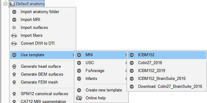
| When you create a new protocol, the program makes a copy of the Colin27 anatomy and sets it as the default for the protocol. It means that you will be able to use the Colin27 brain as a substitute for the subjects without an individual MRI, or as the common brain for group analysis. | Other sets of MRI+surfaces are available to replace the ICBM152/FreeSurfer anatomy. Right-click on ''(Default anatomy)'' > Use template. If a package is not currently available on your system, it will be downloaded from the Brainstorm website and saved in $HOME/.brainstorm/templates. |
| Line 16: | Line 26: |
| Other sets of MRI+surfaces are available to replace the Colin27 anatomy. Right-click on ''(Default anatomy)'' > Use template. If a package is not currently available on your system, it will be downloaded from the Brainstorm website and saved in $HOME/.brainstorm/templates. The available options are: | . {{attachment:changeDefault.gif}} |
| Line 18: | Line 28: |
| * '''Colin27''': Average of 27 scans of the same head, processed with FreeSurfer 5.3: [[http://www.bic.mni.mcgill.ca/ServicesAtlases/Colin27|more information]] * '''Colin27_2012''': Previous version of the default anatomy distributed with Brainstorm * '''ICBM152''': Non-linear average of 152 subjects, processed with FreeSurfer 5.3: [[http://www.bic.mni.mcgill.ca/ServicesAtlases/ICBM152NLin2009|more information]] * '''FSAverage''': Average of 40 subjects using a spherical averaging described in [[http://nmr.mgh.harvard.edu/~fischl/reprints/morphing_human_brain_mapping_reprint.pdf|(Fischl et al. 1999)]].<<BR>>It is the default FreeSurfer brain: please [[https://surfer.nmr.mgh.harvard.edu/registration.html|register here]] if you are using it.<<BR>>More images: http://neuroimage.usc.edu/brainstorm/Tutorials/LabelFreeSurfer#FSAverage_template * '''Infant7w''': 7-week infant brain with the antomical atlas presented in [[http://www.sciencedirect.com/science/article/pii/S105381191400411X|(Kabdebon et al. 2014)]]. |
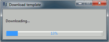
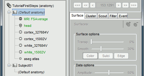
If you click on any of the download options, it downloads it into your templates folder, then the list of files in the (default anatomy) folder is replaced with the new template. . {{attachment:changeDefault2.gif||width="228",height="90"}} {{attachment:changeDefault3.gif||width="352",height="187"}} If the automatic download doesn't work, you can download the templates manually from the [[http://neuroimage.usc.edu/bst/download.php|Download]] page and copy the .zip files directly in the folder $HOME/.brainstorm/templates. == Group analysis == When performing a group analysis with multiple subjects for which you have the individual MRI scans, you need to project the sources estimated on each subject on a common template, as explained in this tutorial: [[Tutorials/CoregisterSubjects|Group analysis]]. For accurate registration between different brains (from a subject to a template or between subjects), you need to use a template that was generated using the '''same program''' as the one you used for running the segmentation of all the subjects of your studies. You can use either '''BrainSuite''' or '''FreeSurfer/''''''CAT12''' for processing the MRIs or your subjects, but you need to use a template that matches this choice in order to use the accurate registration methods. == FreeSurfer templates == Available options: * '''ICBM152''': Distributed directly with the Brainstorm package. Same as ICBM152_2020, but without the white matter envelopes and the volume atlases and SPM registration matrices, in order to minimize the size of the standard Brainstorm distribution. * '''ICBM152_2019''': ICBM 2009c Nonlinear Asymmetric, FreeSurfer 6: [[http://nist.mni.mcgill.ca/?p=904|more info]] * '''Colin27_2016''': Average of 27 scans of the same head, FreeSurfer 5.3: [[http://nist.mni.mcgill.ca/?p=947|more info]] * '''FsAverage_2020''': Average of 40 subjects using a spherical averaging [[http://nmr.mgh.harvard.edu/~fischl/reprints/morphing_human_brain_mapping_reprint.pdf|(Fischl et al. 1999)]].<<BR>>It is the default FreeSurfer brain: please [[https://surfer.nmr.mgh.harvard.edu/registration.html|register here]] if you are using it.<<BR>>More images: http://neuroimage.usc.edu/brainstorm/Tutorials/LabelFreeSurfer#FSAverage_template * '''Oreilly infant templates''': 13 anatomical models for subjects between zero and 24 months of age ([[https://www.biorxiv.org/content/10.1101/2020.06.20.162131v1|O'Reilly et al. 2020]]) |
| Line 27: | Line 55: |
| * Cortex surface: high-resolution (~300.000 vertices) and low-resolution (15.000 vertices) * Head surface: based on the head used for FSAverage in the MNE software * FreeSurfer spherical registration of each hemisphere, with which we can co-register the individual brains processed with FreeSurfer with the selected default anatomy * FreeSurfer surface-based atlases: Desikan-Killiany, Destrieux, Brodman, Mindboggle<<BR>>(plus Yeo2011 and PALS for FSAverage only) |
* Cortex/white surface: high-resolution (~300.000 vertices) and low-resolution (15.000 vertices) * Head layers: scalp, outer skull, inner skull * FreeSurfer spherical registration of each hemisphere, for subject co-registration. * FreeSurfer surface-based atlases: [[https://surfer.nmr.mgh.harvard.edu/fswiki/CorticalParcellation|Desikan-Killiany]], [[https://surfer.nmr.mgh.harvard.edu/fswiki/CorticalParcellation|Destrieux]], [[http://ftp.nmr.mgh.harvard.edu/fswiki/BrodmannAreaMaps|Brodmann]], [[http://www.mindboggle.info/data.html|Mindboggle]]<<BR>>(plus Yeo2011 and PALS for FSAverage only) * ASEG sub-cortical atlas. |
| Line 32: | Line 61: |
| The atlases will be discussed in the following tutorials. For more information on the interactions between FreeSurfer and Brainstorm: [[Tutorials/LabelFreeSurfer|read this tutorial]]. | For more information on the interactions between FreeSurfer and Brainstorm: [[Tutorials/LabelFreeSurfer|read this tutorial]]. |
| Line 34: | Line 63: |
| {{attachment:changeDefault.gif|changeDefault1.gif}} | == BrainSuite templates == Available options: |
| Line 36: | Line 66: |
| If you click on any of the download options, it downloads it into your $HOME/.brainstorm/templates folder: | * '''Colin27_BrainSuite_2016''': Average of 27 scans, processed with BrainSuite 15b: [[http://www.bic.mni.mcgill.ca/ServicesAtlases/Colin27|more information]] * '''ICBM152_BrainSuite_2016''': Non-linear average of 152 subjects, BrainSuite 15b: [[http://www.bic.mni.mcgill.ca/ServicesAtlases/ICBM152NLin2009|more information]] * '''BCI-DNI_BrainSuite_2020''': Single subject atlas from USC, BrainSuite 15c: [[http://brainsuite.org/svreg_atlas_description/|more information]] * '''USCBrain''''''_BrainSuite_2020''': Anatomical and functional hybrid atlas from USC, BrainSuite 17a: [[http://brainsuite.org/uscbrain-description/|more]] |
| Line 38: | Line 71: |
| {{attachment:changeDefault2.gif}} | <<BR>>__Warning__: If you are using '''BrainSuiteAtlas1''' for BrainSuite processing, then you should use '''Colin27_BrainSuite_2016 '''or '''ICBM152_BrainSuite_2016''' as the default anatomy.''' '''If you are using the '''BCI-DNI_brain_atlas''', then you should use '''BCI-DNI_BrainSuite_2016 '''as the template in BrainStorm. |
| Line 40: | Line 73: |
| Then the list of files in the (default anatomy) folder is replaced with the new template. | They all include the following information: |
| Line 42: | Line 75: |
| {{attachment:changeDefault3.gif}} | * T1 MRI volume * Cortex/white surfaces: high-resolution (~300.000 vertices) and low-resolution (15.000 vertices) * Head layers: scalp, outer skull, inner skull * BrainSuite square registration of each hemisphere, for subject co-registration. * BrainSuite surface-based atlas: SVReg For more information on the interactions between BrainSuite and Brainstorm: [[Tutorials/SegBrainSuite|read this tutorial]]. == BrainVISA templates == Note that the BrainVISA-based templates '''do not allow any accurate registration''' procedure. * '''Kabdebon_7w''': 7-week infant brain with the anatomical atlas from [[http://www.sciencedirect.com/science/article/pii/S105381191400411X|(Kabdebon et al. 2014)]]. |
| Line 45: | Line 89: |
| The fiducial points (Nasion, LPA, RPA) used in your recordings might not be the same as the ones used in the anatomy templates in Brainstorm (Colin27, ICBM152, FSAverage). By default, the LPA/RPA points are defined at the junction between the tragus and the helix, as represented with the red dot in the [[http://neuroimage.usc.edu/brainstorm/CoordinateSystems|Coordinates systems page]]. | The fiducial points (Nasion, LPA, RPA) used in your recordings might not be the same as the ones used in the anatomy templates in Brainstorm. By default, the LPA/RPA points are defined at the junction between the tragus and the helix, as represented with the red dot in the [[http://neuroimage.usc.edu/brainstorm/CoordinateSystems|Coordinates systems page]]. |
| Line 47: | Line 91: |
| If you want to use an anatomy template but you are using a different convention when digitizing the position of those points, you have to modify the default positions of the template with the MRI Viewer. | If you want to use an anatomy template but you are using a different convention when digitizing the position of these points, you have to modify the default positions of the template with the MRI Viewer. |
| Line 53: | Line 97: |
== MNI parcellations == |
Using the anatomy templates
Author: Francois Tadel
Brainstorm orients most of its database organization and processing stream for handling anatomical information together with the MEG/EEG recordings. The introduction tutorials start with the import of the T1 MRI of the subject, and this anatomy seems mandatory everywhere. These choices were made because the primary focus of Brainstorm was to estimate brain sources from MEG/EEG, which ideally requires an accurate spatial modelling of the head.
If you don't have access to anatomical images of your subjects or if you are not interested in source reconstruction, Brainstorm will still require that you explicitly define an anatomy; in those case you would use an anatomy template. In the case of group analysis at the source level, you would also use a template on which you would project of the individual results.
Several templates are available, with a large preference for using the MNI ICBM152 package distributed with Brainstorm because it provides the highest level of compatibility between different features within Brainstorm and with other software environments. You might be interested in using another one if you are working with different age ranges, or if you need to obtain results in a specific space. This tutorial provides references to the various templates available in Brainstorm.
Contents
Alternatives
If you do not have access to an individual MR scan of the subject, or if its quality is too low to be processed with FreeSurfer, you have other options:
If you do not have any individual anatomical data: Use the default anatomy
If you have a digitized head shape of the subject: Warp the default anatomy
What changes if you use a default anatomy for EEG or MEG
Changing the default anatomy
When you create a new protocol, the program makes a copy of the ICBM152 anatomy, processed with FreeSurfer 6, and sets it as the default for the protocol. It means that you will be able to use this template brain as a substitute for the subjects without an individual MRI, or as the common brain for group analysis.
Other sets of MRI+surfaces are available to replace the ICBM152/FreeSurfer anatomy. Right-click on (Default anatomy) > Use template. If a package is not currently available on your system, it will be downloaded from the Brainstorm website and saved in $HOME/.brainstorm/templates.
If you click on any of the download options, it downloads it into your templates folder, then the list of files in the (default anatomy) folder is replaced with the new template.
If the automatic download doesn't work, you can download the templates manually from the Download page and copy the .zip files directly in the folder $HOME/.brainstorm/templates.
Group analysis
When performing a group analysis with multiple subjects for which you have the individual MRI scans, you need to project the sources estimated on each subject on a common template, as explained in this tutorial: Group analysis.
For accurate registration between different brains (from a subject to a template or between subjects), you need to use a template that was generated using the same program as the one you used for running the segmentation of all the subjects of your studies.
You can use either BrainSuite or FreeSurfer/CAT12 for processing the MRIs or your subjects, but you need to use a template that matches this choice in order to use the accurate registration methods.
FreeSurfer templates
Available options:
ICBM152: Distributed directly with the Brainstorm package. Same as ICBM152_2020, but without the white matter envelopes and the volume atlases and SPM registration matrices, in order to minimize the size of the standard Brainstorm distribution.
ICBM152_2019: ICBM 2009c Nonlinear Asymmetric, FreeSurfer 6: more info
Colin27_2016: Average of 27 scans of the same head, FreeSurfer 5.3: more info
FsAverage_2020: Average of 40 subjects using a spherical averaging (Fischl et al. 1999).
It is the default FreeSurfer brain: please register here if you are using it.
More images: http://neuroimage.usc.edu/brainstorm/Tutorials/LabelFreeSurfer#FSAverage_templateOreilly infant templates: 13 anatomical models for subjects between zero and 24 months of age (O'Reilly et al. 2020)
They all include the following information:
- T1 MRI volume
- Cortex/white surface: high-resolution (~300.000 vertices) and low-resolution (15.000 vertices)
- Head layers: scalp, outer skull, inner skull
FreeSurfer spherical registration of each hemisphere, for subject co-registration.
FreeSurfer surface-based atlases: Desikan-Killiany, Destrieux, Brodmann, Mindboggle
(plus Yeo2011 and PALS for FSAverage only)- ASEG sub-cortical atlas.
For more information on the interactions between FreeSurfer and Brainstorm: read this tutorial.
BrainSuite templates
Available options:
Colin27_BrainSuite_2016: Average of 27 scans, processed with BrainSuite 15b: more information
ICBM152_BrainSuite_2016: Non-linear average of 152 subjects, BrainSuite 15b: more information
BCI-DNI_BrainSuite_2020: Single subject atlas from USC, BrainSuite 15c: more information
USCBrain_BrainSuite_2020: Anatomical and functional hybrid atlas from USC, BrainSuite 17a: more
Warning: If you are using BrainSuiteAtlas1 for BrainSuite processing, then you should use Colin27_BrainSuite_2016 or ICBM152_BrainSuite_2016 as the default anatomy. If you are using the BCI-DNI_brain_atlas, then you should use BCI-DNI_BrainSuite_2016 as the template in BrainStorm.
They all include the following information:
- T1 MRI volume
- Cortex/white surfaces: high-resolution (~300.000 vertices) and low-resolution (15.000 vertices)
- Head layers: scalp, outer skull, inner skull
BrainSuite square registration of each hemisphere, for subject co-registration.
BrainSuite surface-based atlas: SVReg
For more information on the interactions between BrainSuite and Brainstorm: read this tutorial.
BrainVISA templates
Note that the BrainVISA-based templates do not allow any accurate registration procedure.
Kabdebon_7w: 7-week infant brain with the anatomical atlas from (Kabdebon et al. 2014).
Modify the default MRI fiducials
The fiducial points (Nasion, LPA, RPA) used in your recordings might not be the same as the ones used in the anatomy templates in Brainstorm. By default, the LPA/RPA points are defined at the junction between the tragus and the helix, as represented with the red dot in the Coordinates systems page.
If you want to use an anatomy template but you are using a different convention when digitizing the position of these points, you have to modify the default positions of the template with the MRI Viewer.
- Go to the anatomy view
In (default anatomy), right-click on the MRI > Edit MRI
- Modify the position of the fiducial points to match your own convention
- Click on [Save], it will update the surfaces to match the new coordinate system


