|
Size: 12307
Comment:
|
Size: 17175
Comment:
|
| Deletions are marked like this. | Additions are marked like this. |
| Line 1: | Line 1: |
| = Realistic head model: FEM mesh generation = '''[TUTORIAL UNDER DEVELOPMENT: NOT READY FOR PUBLIC USE]''' |
'''[TUTORIAL UNDER CONSTRUCTION: NOT READY FOR PUBLIC USE]''' |
| Line 4: | Line 3: |
| ''Authors: Takfarinas Medani '' | ---- |
| Line 6: | Line 5: |
| <<TableOfContents(2,2)>> | = FEM mesh generation = ''Authors: [[https://neuroimage.usc.edu/brainstorm/AboutUs/tmedani#preview|Takfarinas Medani]], Francois Tadel'' |
| Line 8: | Line 8: |
| == Introduction == This tutorial present the different method integrated to brainstorm used to generate the FEM mesh. |
FEM forward modeling requires the construction of a 3D model of the head tissues. The volume of the head is divided in small geometrical elements with 4 faces (tetrahedrons) or 6 faces (hexahedrons). Each element is associated with a type of biological tissue (e.g. white matter, gray matter, CSF, skull, skin) and electrical conductivity properties. |
| Line 11: | Line 10: |
| In order to use the finite element method to compute either the EEG/MEG forward problem or the simulate TMS od TDSC stimulation, the mesh of the head model is required. | This page lists the methods integrated with Brainstorm to generate 3D meshes of the head. For a generic introduction to FEM in Brainstorm, refer to the tutorials: [[https://neuroimage.usc.edu/brainstorm/Tutorials/Duneuro|Realistic head model: FEM with DUNEuro]] and [[https://neuroimage.usc.edu/brainstorm/Tutorials/FemMedianNerve|FEM median nerve example]]. |
| Line 13: | Line 12: |
| In this tutorial, we present the different methods available with brainstorm to generate the FEM mesh and how to use them. | <<TableOfContents(3,2)>> |
| Line 15: | Line 14: |
| == FEM Mesh generation methods == The most modern software that are used to generate the volume mesh head model are integrated within brainstorm with an easy graphical interface to use call these tools. |
== Generate FEM mesh == FEM meshes can be computed from surfaces (as the ones generated for the [[https://neuroimage.usc.edu/brainstorm/Tutorials/TutBem#BEM_surfaces|BEM models]]) or from MRI volumes (T1w and/or T2w). The methods that are available when using the popup menu '''Generate FEM mesh''' depend on the selected inputs. |
| Line 18: | Line 17: |
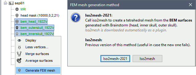
| "Iso2mesh" : This option merges the brainstorm surfaces available on the subject and then generarte the tetrahedral mesh. | === Surfaces === Select a list of surfaces representing the separation between different tissues (holding the CTRL or SHIFT key), then right-click on any of them. The software [[https://neuroimage.usc.edu/brainstorm/Tutorials/FemMesh#Iso2mesh|Iso2mesh]] can create a tetrahedral mesh to represent the tissues between these different layers. |
| Line 20: | Line 20: |
| "Brain2Mesh" : This options uses the MRIs available on the subjects, then it calls the SPM segmentation of the volume into 5 tissus (white, gray, scf, skull and skin). After that it converts into a tetrahedral mesh. | {{attachment:callSurf.gif}} |
| Line 22: | Line 22: |
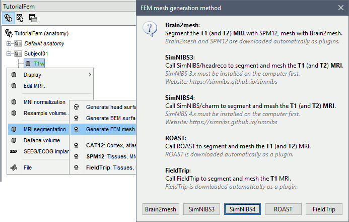
| "SimNibs" : The recommended option, it calls the headreco {ref} and generate a FEM head model | === T1 MRI === Right-click on a T1 MRI available in the databas. Typically, this is the default MRI volume displayed in green in the subject folder. Methods available: [[https://neuroimage.usc.edu/brainstorm/Tutorials/FemMesh#Brain2mesh|Brain2mesh]], [[http://neuroimage.usc.edu/brainstorm/Tutorials/FemMesh#SimNIBS|SimNIBS]], [[http://neuroimage.usc.edu/brainstorm/Tutorials/FemMesh#ROAST|ROAST]], [[http://neuroimage.usc.edu/brainstorm/Tutorials/FemMesh#FieldTrip|FieldTrip]]. |
| Line 24: | Line 25: |
| "FieldTrip" : (in progress) "Roast" : (in progress) | {{attachment:callT1.gif}} |
| Line 26: | Line 27: |
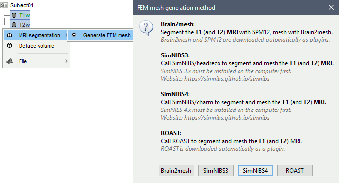
| "headreco" : | === T1+T2 MRI === Select the T1+T2 volumes, then right-click on any of them. The different files are identified based on the tags "T1" and "T2" the file names (as displayed in the Brainstorm database explorer). If these identification tags are not found in the file names, the default MRI (in green) is used as the T1, the other as the T2. If none of the is the default MRI, the first selected file is used as the T1, the second is used as the T2. Methods available: [[https://neuroimage.usc.edu/brainstorm/Tutorials/FemMesh#Brain2mesh|Brain2mesh]], [[http://neuroimage.usc.edu/brainstorm/Tutorials/FemMesh#SimNIBS|SimNIBS]], [[http://neuroimage.usc.edu/brainstorm/Tutorials/FemMesh#ROAST|ROAST]]. |
| Line 28: | Line 30: |
| https://simnibs.github.io/simnibs/build/html/documentation/command_line/headreco.html | {{attachment:callT1T2.gif}} |
| Line 30: | Line 32: |
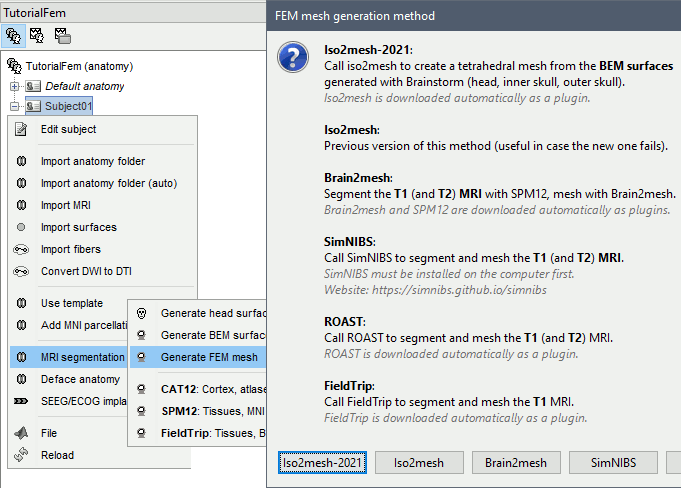
| This function is part of the SimNibs software: | === Anatomy folder === If you right-click on the subject folder > Generate FEM mesh, then Brainstorm offers all the possible options, even the ones that are not applicable to this specific subject. |
| Line 32: | Line 35: |
| Brainstorm integrated a list of the open-source tools that are commonly used by the FEM community to generate either tetra or hexahedra mesh head model either from surface mesh or from MRI. Here is the list of the available methods in brainstorm: | * '''Volume ''''''method''': If using a method based on MRI volumes, the T1 and T2 volumes are detected among all the volumes available based on the tags "T1" and "T2" in the file names (make sure only one file as each of these tags), otherwise use only the default MRI (in green) as the T1. * '''Surface method''': If using a method based on surfaces, the default surfaces (in green) from three categories as selected: the inner skull, the outer skull and the head surfaces. |
| Line 34: | Line 38: |
| - [[http://iso2mesh.sourceforge.net/cgi-bin/index.cgi?Doc/FunctionList|iso2mesh]] | {{attachment:callAnat.gif}} |
| Line 36: | Line 40: |
| - [[http://mcx.space/brain2mesh/|Brain2mesh]] | == Iso2mesh == [[http://iso2mesh.sourceforge.net|Iso2mesh]] is a Matlab/Octave-based mesh generation and processing toolbox, available as a [[https://Tutorials/Plugins|Brainstorm plugin]]. |
| Line 38: | Line 43: |
| - [[https://simnibs.github.io/simnibs/build/html/index.html|SimNibs]] | Brainstorm uses it to generate a FEM tetrahedral mesh from a set of '''nested surfaces''' representing the separation between different tissues of the head. For example, these surfaces can be the ones generated for the computation of a [[https://neuroimage.usc.edu/brainstorm/Tutorials/TutBem#BEM_surfaces|BEM forward model]]. A full example is available in the tutorial [[https://neuroimage.usc.edu/brainstorm/Tutorials/Duneuro#FEM_mesh|Realistic head model: FEM with DUNEuro]]. |
| Line 40: | Line 45: |
| - [[http://www.fieldtriptoolbox.org/|Fieldtrip]] | {{attachment:iso2meshOptions.gif}} |
| Line 42: | Line 47: |
| You can display the full list and a short description by right click on the MRI of the subject and then click the item "Generate FEM Mesh" | ==== Options ==== * '''MergeMesh''': Simply concatenates the input surfaces without any intersection checks. Default option (faster). * '''MergeSurf''': Concatenates and checks for intersections, split intersecting elements. Advanced option (slower). * '''Max tetrahedral volume''': Maximum volume of the tetrahedral element in the mesh. * From our tests, a DUNEuro FEM head model with a value of 0.1 achieves similar results as the OpenMeeg head model computed from the same surfaces. We have also noticed that the result with v = 0,001 is almost similar to v = 0,01. * Increasing the mesh resolution requires more time to generate the mesh, more time and memory to perform the FEM computation and more storage space in the database. * '''Percentage of elements kept''': Parameter between 0-100%, used to keep or not the original input surface nodes. |
| Line 44: | Line 55: |
| {{attachment:meshMethods.JPG||height="400",width="350"}} | ==== Examples ==== * FEM mesh with different values of "Max volume": [10, 1, 0.1, 0.01] - Kept ratio=100%.<<BR>><<BR>> {{attachment:iso2meshMaxvol.gif}} * FEM meshes with only two compartments: This could be useful for investigating the influence of a specific tissue on the EEG/MEG forward solution or on the source localization, or for analyzing SEEG only within the brain volume. On the left: head and outer skull; On the right: inner and outer skull. <<BR>><<BR>> {{attachment:iso2Mesh2layer.gif}} |
| Line 46: | Line 59: |
| == iso2mesh == iso2mesh is a matlab /octave-based mesh generation and processing toolbox. It can [[http://iso2mesh.sourceforge.net/cgi-bin/index.cgi?Doc/Workflow|create 3D tetrahedral finite element (FE) mesh from surfaces, 3D binary and gray-scale volumetric images]] such as segmented MRI/CT scans. |
==== Troubleshooting ==== * '''Tetget failed''': If intersections are present on the surfaces mesh, the iso2mesh FEM mesh generation fails (tetgen). You may need regenerate new surfaces from the MRI. ''' ''' * Alternatively: You may try with the MergSurf option, this option can correct the intersection and create new nodes and elements. However, we do not recommend to use these models for EEG/MEG forward head computations: this is a research topic and it's still under investigation by the FEM communities. == Brain2mesh == [[http://mcx.space/brain2mesh/|Brain2mesh]] is a MATLAB/Octave based 3D mesh generation toolbox dedicated to the creation of high-quality multi-layered brain mesh models. This software is developed by the same team developing Iso2mesh and relies heavily on it. Both are available as a [[https://Tutorials/Plugins|Brainstorm plugins]]. Brainstorm runs the '''SPM12 '''segmentation routine on the '''T1 '''or '''T1+T2 MRI''' volumes to obtain a 5-tissue classifcation (white matter, gray matter, CSF, skull and skin), which is then passed to Brain2mesh for 3D meshing. A full example is available in the tutorial [[https://neuroimage.usc.edu/brainstorm/Tutorials/FemTensors#FEM_mesh|FEM tensors estimation]]. At the moment, Brainstorm can only use the default parameters of Brain2mesh. If you need more options to be available from the interface, please contact us on the user forum. {{attachment:brain2meshCall.gif}} {{attachment:brain2meshMesh.gif}} ==== Troubleshooting ==== * '''SPM-related errors''': If you've been trying multiple methods successively, errors mentioning a spm_*.m function could be due to incompatible versions of SPM12 functions in the Matlab path. Brain2mesh, FieldTrip and ROAST all run different versions of SPM12 from the same instance of Matlab. Solution: '''Restart Matlab''' to get a fresh workspace. == Fieldtrip == - [[http://www.fieldtriptoolbox.org/|Fieldtrip]]: this option calls the volume segmentation function from FieldTrip's pipeline. It generates an hexahedral mesh. This option call [[http://www.fieldtriptoolbox.org/tutorial/headmodel_eeg_fem/|the process of fieldtrip MRI segmentation]] (function ft_volumesegment) and hexahedral mesh generation (ft_meshprepare) develloped by the [[https://www.mrt.uni-jena.de/simbio/index.php?title=Main_Page|SimBio]] team. |
| Line 50: | Line 83: |
| it If iso2mesh is not installed in your computer, Brainstrom will download the last release from this [[https://neuroimage.usc.edu/brainstorm/http://iso2mesh.sourceforge.net/cgi-bin/index.cgi?Download|webpage]] and install it when it'is needed. However, you can also download the iso2mesh from the [[https://github.com/fangq/iso2mesh|github]] and add it to your matlab path. | To use this option, the [[http://www.fieldtriptoolbox.org/getting_started/|Fieldtrip]] and [[https://www.fil.ion.ucl.ac.uk/spm/software/spm12/|SPM toolbox]] should be in your matlab. See the [[https://neuroimage.usc.edu/brainstorm/Tutorials/Plugins|Plugins tutorial]]. |
| Line 53: | Line 86: |
| iso2mesh is used as the basic option by brainstorm to generate FEM mesh from surfaces mesh. | This option can be called by two processes, either from the MRI or from any segmented tissue available on the Brainstorm database. |
| Line 55: | Line 88: |
| Assuming the situation where you have surfaces mesh of your subject available and you have already computed the [[https://neuroimage.usc.edu/brainstorm/Tutorials/TutBem?highlight=(bem)|OpenMeeg]]forward problem. If you want to use the duneuro FEM to compute the forward model, you need to generate the FEM mesh from a similar surface used by OpenMeeg Here is the way to do it : | The mesh generation with the method is faster. It converts all the voxels to hexahedral mesh. |
| Line 57: | Line 90: |
| 1. Richt click on the subject : In this way, brainstorm will load the inner, outer and the head from the subject data. if any of these surfaces is missing, an error will be displayed. 1. Select the 'Generate FEM mesh' item, 1. Select the iso2mesh option, 1. Set the iso2mesh parameters, |
Only the hexahedral mesh is available for this method. You can either call this option from the MRI data or from any segmentation data available on the subject. If you call it from the MRI, a segmentation is processed first, then the mesh. If you call from the tissues, only the mesh process will be performed. |
| Line 62: | Line 92: |
| 1. These options are used by the surf2mesh function. Select either '''MergeMesh''' or '''MergeSurf.''' 1. '''Max tetrahedral volum''' : is the maximum volume of the tetrahedral element in the mesh. '''Pourcentage of the element to keep''': parameter between 0-100%, it used to keep or not the original input surface nodes. |
{{attachment:mriAndTissue.JPG||width="700",height="450"}} |
| Line 66: | Line 94: |
| Also, a full example is explained in this [[https://neuroimage.usc.edu/brainstorm/Duneuro?highlight=(duneuro)|page]]. | Right-click on the MRI (or the tissues), then "Generate FEM Mesh" then select Fieldtrip option. There are two parameters that the user needs to set, the downsampling of the volume and the node shift ratio. |
| Line 68: | Line 96: |
| . | {{attachment:fieldTripMeshCall.jpg||width="650",height="380"}} |
| Line 70: | Line 98: |
| {{attachment:iso2meshProcess.JPG||height="380",width="720"}} | The option "Downsamp volume before meshing" will reduce the number of voxel by this factor. |
| Line 72: | Line 100: |
| Here is a view of mesh obtained with different values of the Max volume = [10, 1, 0.1, 0.01] with a keep ratio = 100%. | The "Shift node" option calls the adaptative mesh generation. The process moves the nodes located on the interface either inward or outward in order to fit the geometry as explained [[http://www.fieldtriptoolbox.org/tutorial/headmodel_eeg_fem/|here]]. This figure shows an example (from Fieldtrip webpage), left the unshifted and on the right the shifted. |
| Line 74: | Line 102: |
| {{attachment:IcbmMeshModels.jpg||height="420",width="700"}} | {{attachment:nodeShiftFigure.JPG||width="500",height="200"}} |
| Line 76: | Line 104: |
| From our experience, a value of 0,1 for the tetrahedral volume achieves similar results as the OpenMeeg computed from the same surfaces. And we have noticed that the result with v = 0,001 is almost similar to v = 0,01. Increasing the mesh resolution needs more time to generate the mesh, more time to perform the FEM computation and of course more memory to store the mesh in the disc. | This method is fast compare to the previous options, the following figures show examples of the mesh obtained with fieldtrip option from the ICBM model. |
| Line 78: | Line 106: |
| If intersections are present on the surfaces mesh, the iso2mesh FEM mesh generation fails (tetgen) and an error will be displayed on the screen. If you face this problem, you need to check the surfaces and/or regenerate new surfaces from the MRI. | {{attachment:fieldTripMeshICBM.JPG||width="700",height="300"}} |
| Line 80: | Line 108: |
| === other use === You can also select any surface mesh, or multiple surfaces (with Shift key), on the brainstorm anatomy windows and then generate tetrahedral mesh y following the same steps above. |
=== Troubleshooting === '''SPM-related errors''': If you've been trying multiple methods successively, errors mentioning a spm_*.m function could be due to incompatible versions of SPM12 functions in the Matlab path. Brain2mesh, FieldTrip and ROAST all run different versions of SPM12 from the same instance of Matlab. Solution: '''Restart Matlab''' to get a fresh workspace. |
| Line 83: | Line 111: |
| Here are some examples using only some 2 tissues. This option could be useful for investigation of tissue influence on the EEG/MEG source localization or analyzing only SEEG within brain volume ... | == SimNIBS == - [[https://simnibs.github.io/simnibs/build/html/index.html|SimNibs]]: this option -recommended for obtaining a realistic model- calls the [[https://simnibs.github.io/simnibs/build/html/documentation/command_line/headreco.html|headreco]] process from SimNIBS toolbox (see the [[https://neuroimage.usc.edu/brainstorm/Tutorials/FemMesh#Additional_Documentation|Additional Documentation]]). It uses the available MRIs for the subject, and then calls SPM and [[http://www.neuro.uni-jena.de/cat/|CAT]] for the segmentation. Then the mesh generation is performed internally by integrated tools (netgen, gmesh and meshfixe). |
| Line 85: | Line 114: |
| {{attachment:otherMesh.JPG||height="320",width="600"}} In this part you can generate your FEM mesh from surfaces that you can get fron the segmentation software (brainSuite, FreeSurfer ....). This process will - merge the surfaces, - check the self intersecting - fixe the size of the mesh - generate the volum mesh - visual checking ... == brain2mesh == Brain2Mesh is a MATLAB/Octave based 3D mesh generation toolbox dedicated to the creation of high-quality multi-layered brain mesh models. |
[[https://simnibs.github.io/simnibs/build/html/index.html|SimNIBS]] software develloped to calculate electric fields caused by Transcranial Electrical Stimulation (TES) and Transcranial Magnetic Stimulation (TMS). From its pipline, Brainstorm integrates the process of the automatic segmentation of MRI images and meshing to create individualized head models. This process is called "headreco and it's explained [[https://simnibs.github.io/simnibs/build/html/documentation/command_line/headreco.html|here]]. |
| Line 105: | Line 117: |
| Brain2Mesh is developed by the same team that developed the iso2mesh toolbox. Therefore iso2mesh is required. So if these toolboxes are not available on your computer, Brainstorm will download the last release and install it when it's needed. | SimNibs is an independent software, Brainstorm call its functions internally therefore you need to install SimNibs and its dependencies. For more details please follow the instructions as explained in [[https://simnibs.github.io/simnibs/build/html/installation/simnibs_installer.html|this webpage]]. |
| Line 107: | Line 119: |
| You may also need the SPM12 toolbox. Brain2mesh is used only to generate tetrahedral mesh from the segmentation output. Therefore a segmentation of the MRI will be performed by SPM when this option is called. | [[https://simnibs.github.io/simnibs/build/html/installation/simnibs_installer.html|Download]] |
| Line 109: | Line 121: |
| More parameters will be added in the next version. If you are using this method you can request our support to help you or to add these parameters asap. | To resume, this process calls SPM12 and CAT for the tissue segmentation, then it calls Gmesh and Netgen for the tetrahedral mesh generation. The mesh is checked and repaired by calling the meshfixe process. Depending on your computer performances, this process will take between 2 to 5 hours. We highly recommend closing all other running processes and applications on your computer in order to speed this process. |
| Line 112: | Line 124: |
| This option is used when you have the individual MRI of the subject either T1 or T1 and T2. As said before, the SPM toolbox is required. The time required for this option is around 1 hour. here is the view of the obtained mesh from a T1 MRI | Brainstorm can call the main function used for the mesh generation frm the main graphical interface. To create individualized models, SimNIBS '''require as''' a T1-weighted image. T2-weighted images are optional, but '''highly recommended'''. The main steps used by SimNIBS are explained in this [[https://simnibs.github.io/simnibs/build/html/tutorial/head_meshing.html|page]]. |
| Line 114: | Line 126: |
| {{attachment:brain2meshModel.JPG||height="450",width="600"}} | When you have your MRI data available on your subject, follow the same steps are explained above, then select the "SimNibs" method. There is one option related to SimNibs, which is the 'Vertex density' or the number of node per mm2 |
| Line 116: | Line 128: |
| More details will be added to this part door. | {{attachment:SimNibsOption.JPG||width="290",height="150"}} |
| Line 118: | Line 130: |
| == Fieldtrip == This option call the process of segmentation and hexa mesh generation of simbio. |
If you have T1 and T2, you need to call this process by a right-click on the subject in order to include the two datasets, or you can select the T1 and T2 then call the Generate FEM mesh process. |
| Line 121: | Line 132: |
| This method calls the function ft_volumesegment then the ft_meshprepare of the field trip. | * If there is an MRI file with the string "T2" in the subject anatomy folder, it will use it |
| Line 123: | Line 134: |
| The mesh generation process is faster. It converts all the voxels to hexahedral mesh. | * Otherwise, if you select explicitly two MRI files with CTRL+Click, it will use the first one as the T1 and the second one as the T2 (this needs to be documented in the tutorial) |
| Line 125: | Line 136: |
| Requirement | The output head models obtained with this method are represented in the following figure. |
| Line 127: | Line 138: |
| The fielstrip toolbox and spm. | {{attachment:SimNibsICBMModel.JPG||width="700",height="300"}} |
| Line 129: | Line 140: |
| Adaptative hexahedral mesh | The model has 5 layers representing the white matter, gray matter, CSF, skull and scalp. |
| Line 131: | Line 142: |
| view of the mesh | == Roast == comming soon under development and integration. |
| Line 133: | Line 145: |
| {{attachment:ernieFieldtrip.JPG||height="450",width="450"}} | For more information, please visit: https://www.parralab.org/roast/ and https://github.com/andypotatohy/roast |
| Line 135: | Line 147: |
| == SimNIBS == === Installation === Please follow the instructions on this [[https://simnibs.github.io/simnibs/build/html/installation/simnibs_installer.html|webapge]]''__ (new brainstorm page that explains how to generate the head model is under development)__'' |
=== Troubleshooting === '''SPM-related errors''': If you've been trying multiple methods successively, errors mentioning a spm_*.m function could be due to incompatible versions of SPM12 functions in the Matlab path. Brain2mesh, FieldTrip and ROAST all run different versions of SPM12 from the same instance of Matlab. Solution: '''Restart Matlab''' to get a fresh workspace. |
| Line 139: | Line 150: |
| in order to do the SimNibs software should be installed on your computer. | == BrainSuite == [[http://brainsuite.org/|BrainSuite]] is a collection of open source software tools that enable largely automated processing of magnetic resonance images (MRI) of the human brain. |
| Line 141: | Line 153: |
| === FEM Head model generation with SimNibs === This method used the SimNibs software. So to call this process, you need to download and install the SimNibs software, the process of the installation is explained in the SimNibs webpage : https://simnibs.github.io/simnibs/build/html/installation/simnibs_installer.html. |
Brainstorm calls BrainSuite tools in order to compute the diffusion tensors from the diffusion wiethed inaging (DWI) data. The diffusion tensor are then converted to conductivity tensors by the linear transformation described by David Tuch et al (ref). |
| Line 144: | Line 155: |
| When you have installed SimNibs, Brainstorm can call the main function used for the mesh generation frm the main graphical interface. Depemding on your computer performances, this process will take between 2 to 5 hours. We highly recommend to close all other running process and application on our computer in order to speed this process. | The conductivity tensors are are associated with the FEM mesh of the head model. Where ecah mesh element have it's own tensors. |
| Line 146: | Line 157: |
| 1- Create new subject within the current protocole | The tensors are used to represnet the anisotropic conductivity of a tissue. The anisotropy means the change on the conductivity by changing the direction. For more information regarding tensors please refers to this page (add link). |
| Line 148: | Line 159: |
| 2- Load the T1 of the subject to the brainstorm database. | === Requirement === BrainSuite is an independant softeware, Brainstoem calls its functions from the core source code, therefore the installation of BrainSuite is required. |
| Line 150: | Line 162: |
| 3- Associate a T2 mri to the subject if it's available (this is better for csf/skull/scalp segmentation) | Please follow the instructions as explained in [[http://forums.brainsuite.org/download/|this webpage]]. |
| Line 152: | Line 164: |
| 4- Right click on the subject, select the "Generate FEM mesh" | Once the instalation is completed, The BrainSuite installation folder must be informed in the Brainstorm preferences (From the Brainsuitrom interface, click on 'File' and then 'Edite preferences'). |
| Line 154: | Line 166: |
| . Select "SIMNIBS", and choose "Tetrahedral element" and keep the other options to the default value. | {{https://user-images.githubusercontent.com/6920058/81406567-1c785400-913a-11ea-9048-28c7459af7da.png|image}} |
| Line 156: | Line 168: |
| The headreco function is fully integrated to brainstorm. With this option, brainstorm can reconstructs a tetrahedral head mesh from T1- and T2-weighted structural MR images. It runs also with only a T1w image, but it will achieve more reliable skull segmentations when a T2w image is supplied. | === When and how to use it === [For this version June 2020, only the tetra mesh are supported and tested.] |
| Line 158: | Line 171: |
| == Tissue anisotropy estimation == === Brainsuite Installation [TODO] === === Volume generation from surface files === |
The BrainSuite pipline is used to estimate the anisotropy of the brain tissues. This process is associated with the [[https://neuroimage.usc.edu/brainstorm/Tutorials/Duneuro?highlight=(duneuro)|DUNEuro FEM computation]]. The conductivity tensors will be assigned to each mesh elements. In order to use this functionnality the DWI data are required. The Niftii files and the assocaited bvec and bval are required. Further more we assume that you have already generated the FEM mesh from the MRI as explained in the previous sections. === Tissue anisotropy estimation === |
| Line 163: | Line 178: |
| === Volume generation from T1/T2 MRI data === You can also generate your own FEM head model and then load it to brainstorm. However the automatic head model generation from from imaging techniques are not accurate and most of the time visual checking are needed and manual correction are required. |
From Brainstorm, BrainSuite is used for the skull stripping, bias field correction and then the diffusion pipline is used to compute the diffusion tensors. |
| Line 166: | Line 180: |
| ==> this depends lagely on the quality of the T1/T2 MRI image(https://simnibs.github.io/simnibs/build/html/tutorial/head_meshing.html). | ref to the maon function : likToGit |
| Line 168: | Line 182: |
| This step is based on the "roast" toolbox (link to roast : https://github.com/andypotatohy/roast | == On the hard drive == Right-click on a FEM mesh > File > View file contents: |
| Line 170: | Line 185: |
| ) that we adapted for the MEEG forward computation. If you want to generate your own FEM head model from an MRI, you will need to download these file (link), then run the bst process as explained here. | {{attachment:femMesh2.gif}} |
| Line 172: | Line 187: |
| * If there is a MRI file with the string "T2" in the subject anatomy folder, it will use it * Otherwise, if you select explicitly two MRI files with CTRL+Click, it will use the first one as the T1 and the second one as the T2 (this needs to be documented in the tutorial) |
TODO |
| Line 175: | Line 189: |
| === FEM Head model template === - Load the FEM volumic mesh (template created from ICBM T1 MRI using SimNibs) |
== Additional Documentation == '''SimNIBS''' |
| Line 178: | Line 192: |
| - Load the surface mesh (template created also from ICBM using ICBM ) and then generates the volume mesh (either tetra or hexa) by calling the tetgen process cia iso2mesh toolbox (if hexa are desired, the tetra mesh will be converted to hexa ... ) | * Website: https://simnibs.github.io/simnibs |
| Line 180: | Line 194: |
| https://github.com/brainstorm-tools/brainstorm3/issues/185#issuecomment-576749612 | * Headreco: https://simnibs.github.io/simnibs/build/html/documentation/command_line/headreco.html |
| Line 182: | Line 196: |
| === Head model based on the level set approach === TODO and Validate == Errors == <<BR>>'''<<TAG(Advanced)>>''' <<BR>> == FEM computation and interface to DUNEuro == === Head model === Number of layers, conductivity value, isotropy/anisotropy/ mesh resolution/ === Electrode model === Check the position of the electrodes and align to the head model (projection if needed) === Source model === Similarly to the spherical and BEM head model, the source position are defined on the cortex surface vertices. We can either define a constrained or not constrained orientation. However, for the FEM model, more paramters could be tuned for the source model. Choice of the source model : PI, Venant, Subtraction, Whitney Panel of the options choice that the user can select. (other wise we will set to default ) '''<<TAG(Advanced)>>''' === Advanced paramaters === - Solver parameters - Electrodes projection - maybe explain here the relevant option of the mini file ?? == Additional documentation == refer to : http://duneuro.org/ https://www.dune-project.org/ https://simnibs.github.io/simnibs/build/html/index.html == Reported Errors & alternative solution == '''<<TAG(Advanced)>>''' simnibs pblm : https://simnibs.github.io/simnibs/build/html/installation/throubleshooting.html == The MEEG forward problem with the FEM == == License == == Reference == == Additional documentation == https://github.com/brainstorm-tools/bst-duneuro/issues/1 https://github.com/brainstorm-tools/brainstorm3/issues/242 |
* Troubleshooting: https://simnibs.github.io/simnibs/build/html/installation/throubleshooting.html |
[TUTORIAL UNDER CONSTRUCTION: NOT READY FOR PUBLIC USE]
FEM mesh generation
Authors: Takfarinas Medani, Francois Tadel
FEM forward modeling requires the construction of a 3D model of the head tissues. The volume of the head is divided in small geometrical elements with 4 faces (tetrahedrons) or 6 faces (hexahedrons). Each element is associated with a type of biological tissue (e.g. white matter, gray matter, CSF, skull, skin) and electrical conductivity properties.
This page lists the methods integrated with Brainstorm to generate 3D meshes of the head. For a generic introduction to FEM in Brainstorm, refer to the tutorials: Realistic head model: FEM with DUNEuro and FEM median nerve example.
Contents
Generate FEM mesh
FEM meshes can be computed from surfaces (as the ones generated for the BEM models) or from MRI volumes (T1w and/or T2w). The methods that are available when using the popup menu Generate FEM mesh depend on the selected inputs.
Surfaces
Select a list of surfaces representing the separation between different tissues (holding the CTRL or SHIFT key), then right-click on any of them. The software Iso2mesh can create a tetrahedral mesh to represent the tissues between these different layers.
T1 MRI
Right-click on a T1 MRI available in the databas. Typically, this is the default MRI volume displayed in green in the subject folder. Methods available: Brain2mesh, SimNIBS, ROAST, FieldTrip.
T1+T2 MRI
Select the T1+T2 volumes, then right-click on any of them. The different files are identified based on the tags "T1" and "T2" the file names (as displayed in the Brainstorm database explorer). If these identification tags are not found in the file names, the default MRI (in green) is used as the T1, the other as the T2. If none of the is the default MRI, the first selected file is used as the T1, the second is used as the T2. Methods available: Brain2mesh, SimNIBS, ROAST.
Anatomy folder
If you right-click on the subject folder > Generate FEM mesh, then Brainstorm offers all the possible options, even the ones that are not applicable to this specific subject.
Volume method: If using a method based on MRI volumes, the T1 and T2 volumes are detected among all the volumes available based on the tags "T1" and "T2" in the file names (make sure only one file as each of these tags), otherwise use only the default MRI (in green) as the T1.
Surface method: If using a method based on surfaces, the default surfaces (in green) from three categories as selected: the inner skull, the outer skull and the head surfaces.
Iso2mesh
Iso2mesh is a Matlab/Octave-based mesh generation and processing toolbox, available as a Brainstorm plugin.
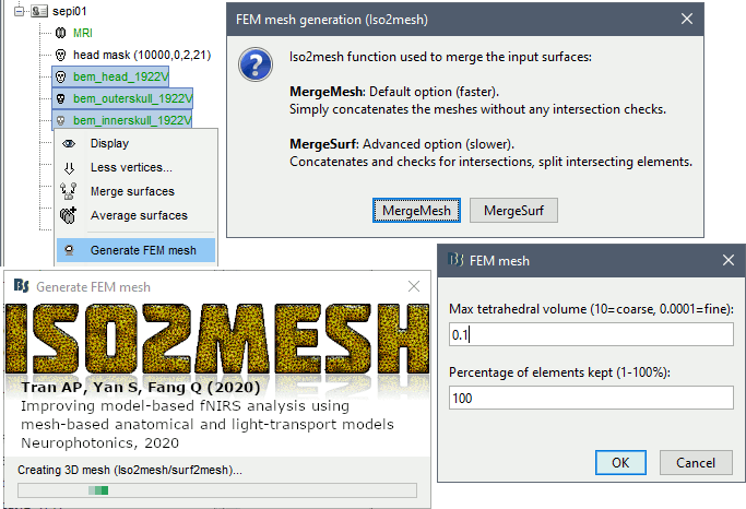
Brainstorm uses it to generate a FEM tetrahedral mesh from a set of nested surfaces representing the separation between different tissues of the head. For example, these surfaces can be the ones generated for the computation of a BEM forward model. A full example is available in the tutorial Realistic head model: FEM with DUNEuro.
Options
MergeMesh: Simply concatenates the input surfaces without any intersection checks. Default option (faster).
MergeSurf: Concatenates and checks for intersections, split intersecting elements. Advanced option (slower).
Max tetrahedral volume: Maximum volume of the tetrahedral element in the mesh.
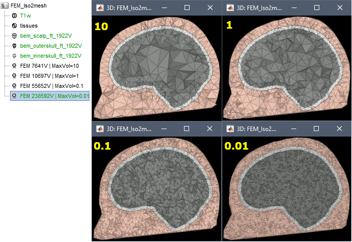
From our tests, a DUNEuro FEM head model with a value of 0.1 achieves similar results as the OpenMeeg head model computed from the same surfaces. We have also noticed that the result with v = 0,001 is almost similar to v = 0,01.
- Increasing the mesh resolution requires more time to generate the mesh, more time and memory to perform the FEM computation and more storage space in the database.
Percentage of elements kept: Parameter between 0-100%, used to keep or not the original input surface nodes.
Examples
FEM mesh with different values of "Max volume": [10, 1, 0.1, 0.01] - Kept ratio=100%.
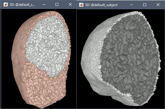
FEM meshes with only two compartments: This could be useful for investigating the influence of a specific tissue on the EEG/MEG forward solution or on the source localization, or for analyzing SEEG only within the brain volume. On the left: head and outer skull; On the right: inner and outer skull.
Troubleshooting
Tetget failed: If intersections are present on the surfaces mesh, the iso2mesh FEM mesh generation fails (tetgen). You may need regenerate new surfaces from the MRI.
Alternatively: You may try with the MergSurf option, this option can correct the intersection and create new nodes and elements. However, we do not recommend to use these models for EEG/MEG forward head computations: this is a research topic and it's still under investigation by the FEM communities.
Brain2mesh
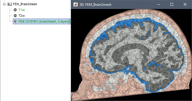
Brain2mesh is a MATLAB/Octave based 3D mesh generation toolbox dedicated to the creation of high-quality multi-layered brain mesh models. This software is developed by the same team developing Iso2mesh and relies heavily on it. Both are available as a Brainstorm plugins.
Brainstorm runs the SPM12 segmentation routine on the T1 or T1+T2 MRI volumes to obtain a 5-tissue classifcation (white matter, gray matter, CSF, skull and skin), which is then passed to Brain2mesh for 3D meshing. A full example is available in the tutorial FEM tensors estimation.
At the moment, Brainstorm can only use the default parameters of Brain2mesh. If you need more options to be available from the interface, please contact us on the user forum.

Troubleshooting
SPM-related errors: If you've been trying multiple methods successively, errors mentioning a spm_*.m function could be due to incompatible versions of SPM12 functions in the Matlab path. Brain2mesh, FieldTrip and ROAST all run different versions of SPM12 from the same instance of Matlab. Solution: Restart Matlab to get a fresh workspace.
Fieldtrip
- Fieldtrip: this option calls the volume segmentation function from FieldTrip's pipeline. It generates an hexahedral mesh.
This option call the process of fieldtrip MRI segmentation (function ft_volumesegment) and hexahedral mesh generation (ft_meshprepare) develloped by the SimBio team.
Requirement
To use this option, the Fieldtrip and SPM toolbox should be in your matlab. See the Plugins tutorial.
When and how to use it
This option can be called by two processes, either from the MRI or from any segmented tissue available on the Brainstorm database.
The mesh generation with the method is faster. It converts all the voxels to hexahedral mesh.
Only the hexahedral mesh is available for this method. You can either call this option from the MRI data or from any segmentation data available on the subject. If you call it from the MRI, a segmentation is processed first, then the mesh. If you call from the tissues, only the mesh process will be performed.
Right-click on the MRI (or the tissues), then "Generate FEM Mesh" then select Fieldtrip option. There are two parameters that the user needs to set, the downsampling of the volume and the node shift ratio.
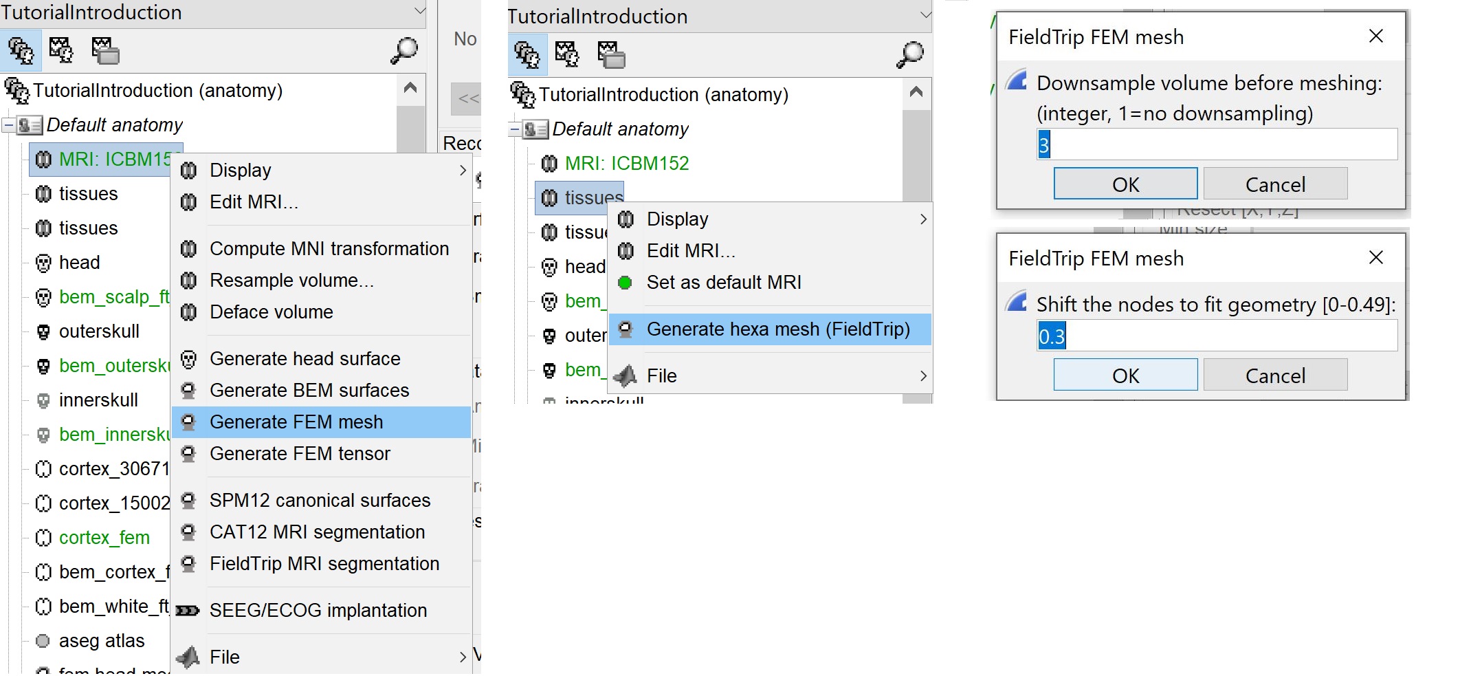
The option "Downsamp volume before meshing" will reduce the number of voxel by this factor.
The "Shift node" option calls the adaptative mesh generation. The process moves the nodes located on the interface either inward or outward in order to fit the geometry as explained here. This figure shows an example (from Fieldtrip webpage), left the unshifted and on the right the shifted.
This method is fast compare to the previous options, the following figures show examples of the mesh obtained with fieldtrip option from the ICBM model.
Troubleshooting
SPM-related errors: If you've been trying multiple methods successively, errors mentioning a spm_*.m function could be due to incompatible versions of SPM12 functions in the Matlab path. Brain2mesh, FieldTrip and ROAST all run different versions of SPM12 from the same instance of Matlab. Solution: Restart Matlab to get a fresh workspace.
SimNIBS
- SimNibs: this option -recommended for obtaining a realistic model- calls the headreco process from SimNIBS toolbox (see the Additional Documentation). It uses the available MRIs for the subject, and then calls SPM and CAT for the segmentation. Then the mesh generation is performed internally by integrated tools (netgen, gmesh and meshfixe).
SimNIBS software develloped to calculate electric fields caused by Transcranial Electrical Stimulation (TES) and Transcranial Magnetic Stimulation (TMS). From its pipline, Brainstorm integrates the process of the automatic segmentation of MRI images and meshing to create individualized head models. This process is called "headreco and it's explained here.
Requirement
SimNibs is an independent software, Brainstorm call its functions internally therefore you need to install SimNibs and its dependencies. For more details please follow the instructions as explained in this webpage.
To resume, this process calls SPM12 and CAT for the tissue segmentation, then it calls Gmesh and Netgen for the tetrahedral mesh generation. The mesh is checked and repaired by calling the meshfixe process. Depending on your computer performances, this process will take between 2 to 5 hours. We highly recommend closing all other running processes and applications on your computer in order to speed this process.
When and how to use it
Brainstorm can call the main function used for the mesh generation frm the main graphical interface. To create individualized models, SimNIBS require as a T1-weighted image. T2-weighted images are optional, but highly recommended. The main steps used by SimNIBS are explained in this page.
When you have your MRI data available on your subject, follow the same steps are explained above, then select the "SimNibs" method. There is one option related to SimNibs, which is the 'Vertex density' or the number of node per mm2
If you have T1 and T2, you need to call this process by a right-click on the subject in order to include the two datasets, or you can select the T1 and T2 then call the Generate FEM mesh process.
* If there is an MRI file with the string "T2" in the subject anatomy folder, it will use it
- Otherwise, if you select explicitly two MRI files with CTRL+Click, it will use the first one as the T1 and the second one as the T2 (this needs to be documented in the tutorial)
The output head models obtained with this method are represented in the following figure.
The model has 5 layers representing the white matter, gray matter, CSF, skull and scalp.
Roast
comming soon under development and integration.
For more information, please visit: https://www.parralab.org/roast/ and https://github.com/andypotatohy/roast
Troubleshooting
SPM-related errors: If you've been trying multiple methods successively, errors mentioning a spm_*.m function could be due to incompatible versions of SPM12 functions in the Matlab path. Brain2mesh, FieldTrip and ROAST all run different versions of SPM12 from the same instance of Matlab. Solution: Restart Matlab to get a fresh workspace.
BrainSuite
BrainSuite is a collection of open source software tools that enable largely automated processing of magnetic resonance images (MRI) of the human brain.
Brainstorm calls BrainSuite tools in order to compute the diffusion tensors from the diffusion wiethed inaging (DWI) data. The diffusion tensor are then converted to conductivity tensors by the linear transformation described by David Tuch et al (ref).
The conductivity tensors are are associated with the FEM mesh of the head model. Where ecah mesh element have it's own tensors.
The tensors are used to represnet the anisotropic conductivity of a tissue. The anisotropy means the change on the conductivity by changing the direction. For more information regarding tensors please refers to this page (add link).
Requirement
BrainSuite is an independant softeware, Brainstoem calls its functions from the core source code, therefore the installation of BrainSuite is required.
Please follow the instructions as explained in this webpage.
Once the instalation is completed, The BrainSuite installation folder must be informed in the Brainstorm preferences (From the Brainsuitrom interface, click on 'File' and then 'Edite preferences').

When and how to use it
[For this version June 2020, only the tetra mesh are supported and tested.]
The BrainSuite pipline is used to estimate the anisotropy of the brain tissues. This process is associated with the DUNEuro FEM computation. The conductivity tensors will be assigned to each mesh elements.
In order to use this functionnality the DWI data are required. The Niftii files and the assocaited bvec and bval are required. Further more we assume that you have already generated the FEM mesh from the MRI as explained in the previous sections.
Tissue anisotropy estimation
From Brainstorm, BrainSuite is used for the skull stripping, bias field correction and then the diffusion pipline is used to compute the diffusion tensors.
ref to the maon function : likToGit
On the hard drive
Right-click on a FEM mesh > File > View file contents:
TODO
Additional Documentation
SimNIBS
